Alternate header for print version


Licensing
Public Domain: This image is in the public domain and thus free of any copyright restrictions. However, as is the norm in scientific publishing and as a matter of courtesy, any user should credit the content provider for any public or private use of this image whenever possible. Learn more
tweet  
*CIL – Cell Image Library accession number. Please use this to reference an image.

CIL:35388*  Cite 
Description

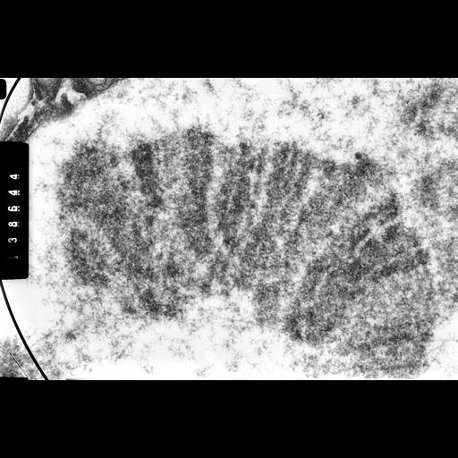
High Voltage (1MeV) TEM image of a 'thick' section of polytene chromosome from salivary gland of 3rd instar larva of Drosophila melanogaster showing the band-interband organization. This image was captured with a specimen tilt of 56 degrees. Grouped with it is an image of the same region taken at a tilt angle of 45 degrees, providing an oblique stereo view of the structure.

Technical Details

Salivary glands were dissected, fixed with paraformaldehyde, post-fixed with osmium tetroxide, and embedded in plastic. 4 micron sections were cut, stained with aqueous uranyl magnesium acetate, and observed with the Wisconsin HVEM at 1 MeV. See Ris, H 1981 Stereoscopic Electron Microscopy of chromosomes. Meth Cell Biol 22:77-96; Ris, H 1978 Preparation of chromatin and chromosomes for electron microscopy. Meth Cell Biol 18:229-246

Biological Sources
NCBI Organism Classification
Drosophila melanogaster
Cell Type
salivary gland
Cellular Component
polytene chromosome
Biological Context
Biological Process
chromosome organization
Attribution
Names
Hans Ris, University of Wisconsin, Madison, WI
Citation
Digital Object Identifier (DOI)
doi:10.7295/W9CIL35388
Archival Resource Key (ARK)
ark:/b7295/w9cil35388
Grouping This image is part of a group.
Imaging
Image Type
film
Image Mode
Parameters Imaged
electron density
Source of Contrast
differences in adsorption or binding of stain
Visualization Methods
uranyl salt
Processing History
unprocessed raw data
Sample Preparation
Methods
chemically fixed tissue
paraformaldehyde fixation
osmium tetroxide fixed tissue
tissue in plastic embedment
Relation To Intact Cell
microtome-sectioned tissue
Dimensions
Spatial Axis Image Size Pixel Size
Y 3690px 1.5nm