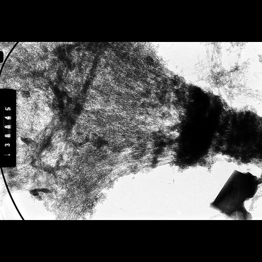
High Voltage (1MeV) TEM image of a squash preparation of polytene chromosomes from salivary gland of 3rd instar larva of Drosophila melanogaster showing the band-interband organization. This image was captured with a specimen tilt of 45 degrees. Grouped with it is an image of the same region taken at a tilt angle of 55 degrees, providing an oblique stereo view of the structure.
Salivary glands were dissected, exposed to 0.07 M KCl, fixed in 3:1 acetic acid:ethanol and squashed in 50% acetic acid. Slides were frozen in liquid nitrogen, the cover slip removed, and the slide immersed in 1% uranyl acetate for 5 min followed by dehydration in ethanol and transfer to amyl acetate. 2% parlodion in amyl acetate was spread over the slide and allowed to air dry. Areas for observation were scored, released by water immersion, picked up on formvar-coated grids and observed with the Wisconsin HVEM at 1 MeV. See Ris, H 1981 Stereoscopic Electron Microscopy of chromosomes. Meth Cell Biol 22:77-96; Ris, H 1978 Preparation of chromatin and chromosomes for electron microscopy. Meth Cell Biol 18:229-246.
| Spatial Axis | Image Size | Pixel Size |
|---|---|---|
| X | 5600px | 1.5nm |
| Y | 3817px | 1.5nm |