Alternate header for print version


Licensing
Public Domain: This image is in the public domain and thus free of any copyright restrictions. However, as is the norm in scientific publishing and as a matter of courtesy, any user should credit the content provider for any public or private use of this image whenever possible. Learn more
tweet  
*CIL – Cell Image Library accession number. Please use this to reference an image.

CIL:35452*  Cite 
Description

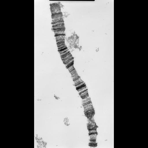
Low magnification of whole polytene chromosome from 3rd instar larva of Drosophila melanogaster imaged with high voltage EM at 1MeV showing the characteristic pattern of bands and interbands.

Technical Details

Salivary gland cells were squashed in 45% acetic acid, slides frozen in liquid nitrogen, cover slips removed, stained with uranyl magnesium acetate, dehydrated with ethanol, transferred to formvar coated grids, critical point dried, and imaged at 1MeV with the Wisconsin HVEM. See Ris (1978) Preparation of chromatin and chromosomes for electron microscopy. Meth Cell Biol 18:229-246; Ris (1981) Stereoscopic microscopy of chromosomes. Meth Cell Biol 22:77-96.

Biological Sources
NCBI Organism Classification
Drosophila melanocephala
Cell Type
salivary gland
Cellular Component
polytene chromosome
Attribution
Names
Hans Ris
Citation
Digital Object Identifier (DOI)
doi:10.7295/W9CIL35452
Archival Resource Key (ARK)
ark:/b7295/w9cil35452
Imaging
Image Type
film
Image Mode
Parameters Imaged
electron density
Source of Contrast
differences in adsorption or binding of stain
Visualization Methods
uranyl salt
Processing History
unprocessed raw data
Sample Preparation
Methods
coagulating-fixative fixed tissue
Relation To Intact Cell
whole mounted tissue
Dimensions
Spatial Axis Image Size Pixel Size
X 3750px 2.3µm
Y 6941px 2.3µm