Alternate header for print version


Licensing
Public Domain: This image is in the public domain and thus free of any copyright restrictions. However, as is the norm in scientific publishing and as a matter of courtesy, any user should credit the content provider for any public or private use of this image whenever possible. Learn more
tweet  
*CIL – Cell Image Library accession number. Please use this to reference an image.

CIL:35483*  Cite 
Description

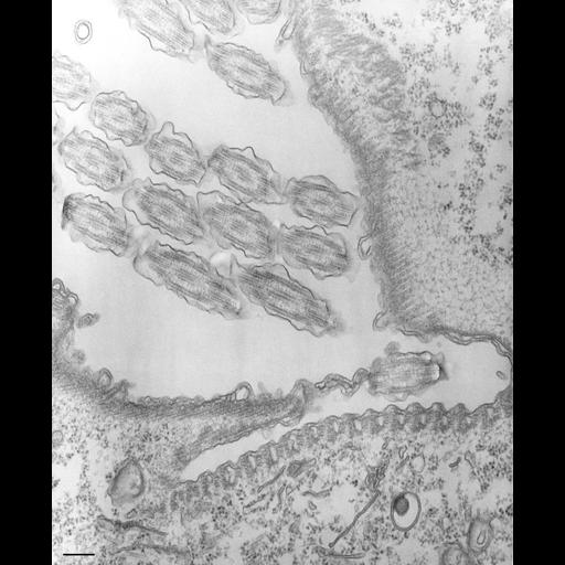
An image of the oral ribs with their microtubular ribbons and alveolar tubes pass through the cytostome to the cytopharynx and nascent vacuole region. In this view the left wall of the buccal cavity is in grazing section so the partial layer of microtubules appears on both sides of the cytostome. The only membrane not associated with alveoli is that at the beginning of the cytopharynx. TEM taken on 8/29/67 by R. Allen with Philips 200 operating at 60kV. Neg. 25,700X. Bar = 0.5µm. A print of the negative was scanned and processed in Photoshop. This image is best used for qualitative analysis. A high resolution image (CIL:34847) is available for quantitative analysis.

Technical Details

Standard glutaraldehyde fixation followed by osmium tetroxide, dehydrated in alcohol and embedded in an epoxy resin. Microtome sections prepared at approximately 75nm thickness. Additional information available at (http://www5.pbrc.hawaii.edu/allen/).

Biological Sources
NCBI Organism Classification
Tetrahymena pyriformis
Cell Type
cell by organism
eukaryotic cell
Eukaryotic Protist
Ciliated Protist
Cellular Component
oral apparatus
Biological Context
Biological Process
oral apparatus organization
Attribution
Names
Richard Allen (University of Hawaii)
Citation
Digital Object Identifier (DOI)
doi:10.7295/W9CIL35483
Archival Resource Key (ARK)
ark:/b7295/w9cil35483
Grouping This image is part of a group.
Imaging
Image Type
transmission electron microscopy (TEM)
illumination by electrons
Image Mode
detection of electrons
film
Parameters Imaged
electron density
Source of Contrast
stain with broad specificity
Visualization Methods
stain with broad specificity
osmium tetroxide
uranyl salt
lead salt
Processing History
recorded image
film
a print was scanned for Photoshop
Data Qualifiers
processed data
Dimensions
Spatial Axis Image Size Pixel Size
X 3208px ——
Y 3924px ——