Alternate header for print version


Licensing
Attribution Non-Commercial; No Derivatives:This image is licensed under a Creative Commons Attribution, Non-Commercial, No Derivatives License. View License Deed | View Legal Code
tweet  
*CIL – Cell Image Library accession number. Please use this to reference an image.

CIL:35964*  Cite 
Description

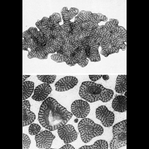
Figure 346 from Chapter 14 (Sperm Flagellum) of 'The Cell, 2nd Ed.' by Don W. Fawcett M.D. Top panel: A cross section of sperm tail of the cecidomyid fly, Diplolaboncus tumorificus shows mitochondria scattered among doublets of microtubules. Lower panel, sperm tail organization in a related species of fly shows about 100 doublets a spiral or row-like arrangement. Figure from Baccetti and Dallai, J. Ultrastr. Res. 55: 50-69, 1976. A PDF copy of the accompanying chapter is available on the ASCB’s BioEDUCATE website.

Biological Sources
NCBI Organism Classification
Cecidomyiinae
Cell Type
sperm
Cellular Component
microtubule-based flagellum
Biological Context
Biological Process
fertilization
Attribution
Names
Don W. Fawcett
B. Bacetti; R. Dallai
Published
ISBN 0721635849
Link
http://www.ascb.org/bioeducate/FawcettTheCell...
Citation
Digital Object Identifier (DOI)
doi:10.7295/W9CIL35964
Archival Resource Key (ARK)
ark:/b7295/w9cil35964
Grouping This image is part of a group.
Imaging
Image Type
transmission electron microscopy (TEM)
Image Mode
illumination by electrons
detection of electrons
Parameters Imaged
electron density
Source of Contrast
stain with broad specificity
Visualization Methods
osmium tetroxide
Data Qualifiers
processed data
Dimensions
Spatial Axis Image Size Pixel Size
X 876px ——
Y 1252px ——