Alternate header for print version


Licensing
Attribution Non-Commercial; No Derivatives:This image is licensed under a Creative Commons Attribution, Non-Commercial, No Derivatives License. View License Deed | View Legal Code
tweet  
*CIL – Cell Image Library accession number. Please use this to reference an image.

CIL:35965*  Cite 
Description

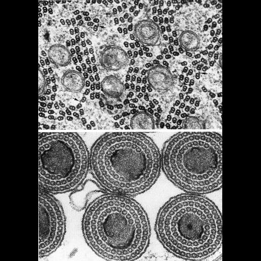
Figures 347 (upper) and 348 (lower) from Chapter 14 (Sperm Flagellum) of 'The Cell, 2nd Ed.' by Don W. Fawcett M.D. Sperm tail organization in some insects deviates from the organization observed in mammals. Upper: A cross section of sperm tail of the cecidomyid fly, Diplolaboncus tumorificus shows only an outer dynein arm (arrows) emerging from doublets of microtubules. Lower: Transverse sections through spermatozoa of the oyster shell scale insect, Lipidosaphes, shows singlet microtubules aranged in concentric rings around a central core. Upper micrograph courtesy for Romao Dallai; lower from Phillips, Spermiogenesis, Academic Press, New York, 1974. A PDF copy of the accompanying chapter is available on the ASCB’s BioEDUCATE website.

Biological Sources
NCBI Organism Classification
Cecidomyiinae
Lepidosaphes
Cell Type
sperm
Cellular Component
microtubule-based flagellum
Biological Context
Biological Process
fertilization
Attribution
Names
Don W. Fawcett
Romano Dallai
David Phillips
Published
ISBN 0721635849
Link
http://www.ascb.org/bioeducate/FawcettTheCell...
Citation
Digital Object Identifier (DOI)
doi:10.7295/W9CIL35965
Archival Resource Key (ARK)
ark:/b7295/w9cil35965
Grouping This image is part of a group.
Imaging
Image Type
transmission electron microscopy (TEM)
Image Mode
illumination by electrons
detection of electrons
Parameters Imaged
electron density
Source of Contrast
stain with broad specificity
Visualization Methods
osmium tetroxide
Data Qualifiers
processed data
Dimensions
Spatial Axis Image Size Pixel Size
X 886px ——
Y 1258px ——