Alternate header for print version


Licensing
Attribution Non-Commercial; No Derivatives:This image is licensed under a Creative Commons Attribution, Non-Commercial, No Derivatives License. View License Deed | View Legal Code
tweet  
*CIL – Cell Image Library accession number. Please use this to reference an image.

CIL:35989*  Cite 
Description

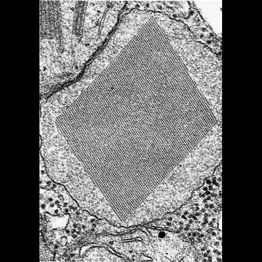
Figure 368 from Chapter 15 (Cytoplasmic Inclusions) of 'The Cell, 2nd Ed.' by Don W. Fawcett M.D. A microbody or peroxisome from a cell in a leaf of tobacco (Nicotiana tabacum). Crystals within microbodies, or peroxisomes, are common, and these organelles appear in plant, as well as animal, cells. Image from Frederick, S.E., Newcomb, E.H., Science 163:1353, 1969; copyright held by AAAS. A PDF copy of the accompanying chapter is available on the ASCB’s BioEDUCATE website.

Biological Sources
NCBI Organism Classification
Nicotiana tabacum
Cell Type
plant cell
Cellular Component
microbody
peroxisome
Attribution
Names
Don W. Fawcett
S.E. Frederick; E.H. Newcomb
Published
ISBN 0721635849
Link
http://www.ascb.org/bioeducate/FawcettTheCell...
Citation
Digital Object Identifier (DOI)
doi:10.7295/W9CIL35989
Archival Resource Key (ARK)
ark:/b7295/w9cil35989
Grouping This image is part of a group.
Imaging
Image Type
transmission electron microscopy (TEM)
Image Mode
illumination by electrons
detection of electrons
Parameters Imaged
electron density
Source of Contrast
stain with broad specificity
Visualization Methods
osmium tetroxide
Data Qualifiers
processed data
Dimensions
Spatial Axis Image Size Pixel Size
X 888px ——
Y 1272px ——