Alternate header for print version


Licensing
Attribution Non-Commercial; No Derivatives:This image is licensed under a Creative Commons Attribution, Non-Commercial, No Derivatives License. View License Deed | View Legal Code
tweet  
*CIL – Cell Image Library accession number. Please use this to reference an image.

CIL:35993*  Cite 
Description

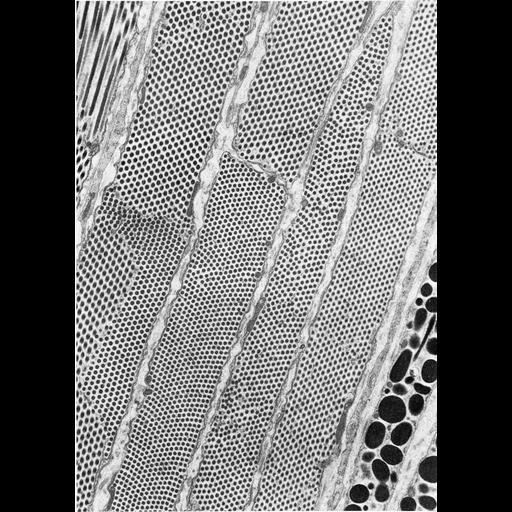
Figure 372 from Chapter 15 (Cytoplasmic Inclusions) of 'The Cell, 2nd Ed.' by Don W. Fawcett M.D. In the tapetum lucidum of a cat's eye, the cytoplasm of cells is filled with highly ordered rod-like crystals containing a compound rich in zinc and cysteine. Mitochondria and other organelles are displaced to the periphery of the cell. At the lower right are portions of two melanin-containing pigment cells. Image by Giuseppina Raviola. A PDF copy of the accompanying chapter is available on the ASCB’s BioEDUCATE website.

Biological Sources
NCBI Organism Classification
Felis catus
Cellular Component
inclusion body
Biological Context
Biological Process
detection of light stimulus involved in visual perception
Attribution
Names
Don W. Fawcett
Giuseppina Raviola
Published
ISBN 0721635849
Link
http://www.ascb.org/bioeducate/FawcettTheCell...
Citation
Digital Object Identifier (DOI)
doi:10.7295/W9CIL35993
Archival Resource Key (ARK)
ark:/b7295/w9cil35993
Grouping This image is part of a group.
Imaging
Image Type
transmission electron microscopy (TEM)
Image Mode
illumination by electrons
detection of electrons
Parameters Imaged
electron density
Source of Contrast
stain with broad specificity
Visualization Methods
osmium tetroxide
Data Qualifiers
processed data
Dimensions
Spatial Axis Image Size Pixel Size
X 897px ——
Y 1269px ——