Alternate header for print version


Licensing
Attribution Non-Commercial; No Derivatives:This image is licensed under a Creative Commons Attribution, Non-Commercial, No Derivatives License. View License Deed | View Legal Code
tweet  
*CIL – Cell Image Library accession number. Please use this to reference an image.

CIL:35999*  Cite 
Description

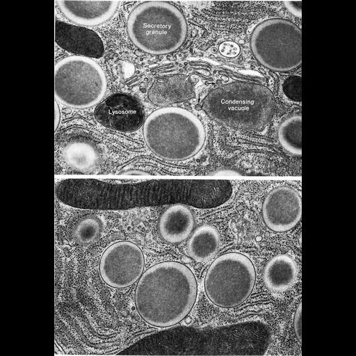
Figures 379 (upper) and 380 (lower) from Chapter 15 (Cytoplasmic Inclusions) of 'The Cell, 2nd Ed.' by Don W. Fawcett M.D. Freeze substitution preparations of gerbil parotid gland show condensing vacuoles arising from the Golgi (arrows, upper panel). Both upper and lower figures show secretory granules with a dense core and lighter cortex. Image by Atsuchi Ichikawa. A PDF copy of the accompanying chapter is available on the ASCB’s BioEDUCATE website.

Biological Sources
NCBI Organism Classification
Gerbillinae
Cell Type
secretory cell
Cellular Component
secretory granule
lysosome
Golgi apparatus
endoplasmic reticulum
mitochondrion
Biological Context
Biological Process
secretory granule organization
Attribution
Names
Don W. Fawcett
Atsuchi Ichikawa
Published
ISBN 0721635849
Link
http://www.ascb.org/bioeducate/FawcettTheCell...
Citation
Digital Object Identifier (DOI)
doi:10.7295/W9CIL35999
Archival Resource Key (ARK)
ark:/b7295/w9cil35999
Grouping This image is part of a group.
Imaging
Image Type
transmission electron microscopy (TEM)
Image Mode
illumination by electrons
detection of electrons
Parameters Imaged
electron density
Source of Contrast
stain with broad specificity
Visualization Methods
osmium tetroxide
Data Qualifiers
processed data
Dimensions
Spatial Axis Image Size Pixel Size
X 885px ——
Y 1273px ——