Alternate header for print version


Licensing
Attribution Non-Commercial; No Derivatives:This image is licensed under a Creative Commons Attribution, Non-Commercial, No Derivatives License. View License Deed | View Legal Code
tweet  
*CIL – Cell Image Library accession number. Please use this to reference an image.

CIL:36016*  Cite 
Description

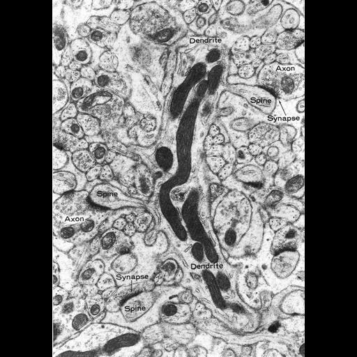
Figure 398 from Chapter 15 (Cytoplasmic Inclusions) of 'The Cell, 2nd Ed.' by Don W. Fawcett M.D. Excitatory synapses on dendritic spines of a Purkinje cell from the rat cerebellum. Axon terminals from parallel fibers can be seen making synaptic contacts on the heads of the spines. Image from Peters, Palay and Webster, Fine Structure of the Nervous System, Philadelphia, W.B. Saunders Co. 1976. A PDF copy of the accompanying chapter is available on the ASCB’s BioEDUCATE website.

Biological Context
Biological Process
synapse organization
Attribution
Names
Don W. Fawcett
Alan Peters; Sanford Palay; Henry Webster
Published
ISBN 0721635849
Link
http://www.ascb.org/bioeducate/FawcettTheCell...
Citation
Digital Object Identifier (DOI)
doi:10.7295/W9CIL36016
Archival Resource Key (ARK)
ark:/b7295/w9cil36016
Grouping This image is part of a group.
Imaging
Image Type
transmission electron microscopy (TEM)
Image Mode
illumination by electrons
detection of electrons
Parameters Imaged
electron density
Source of Contrast
stain with broad specificity
Visualization Methods
osmium tetroxide
Data Qualifiers
processed data
Dimensions
Spatial Axis Image Size Pixel Size
X 889px ——
Y 1257px ——