Alternate header for print version


Licensing
Attribution Non-Commercial; No Derivatives:This image is licensed under a Creative Commons Attribution, Non-Commercial, No Derivatives License. View License Deed | View Legal Code
tweet  
*CIL – Cell Image Library accession number. Please use this to reference an image.

CIL:36068*  Cite 
Description

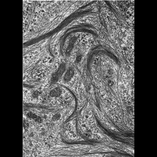
Figure 436 from Chapter 16 (Cytoplasmic matrix and cytoskeleton) of 'The Cell, 2nd Ed.' by Don W. Fawcett M.D. Cytoplasm of a Sertoli cell from testis of the ground squirrel, Citellus lateralis in culture. When grown in vitro, actin filaments tend to aggregate into coarse bundles. Large aggregations of parallel filaments correspond to stress fibers. Image by don Vogl. A PDF copy of the accompanying chapter is available on the ASCB’s BioEDUCATE website.

Biological Sources
NCBI Organism Classification
Spermophilus citellus
Cell Type
Sertoli cell
Cellular Component
actin filament
actin filament bundle
stress fiber
Biological Context
Biological Process
cell motility
cell adhesion
Attribution
Names
Don W. Fawcett
Wayne Vogl
Published
ISBN 0721635849
Link
http://www.ascb.org/bioeducate/FawcettTheCell...
Citation
Digital Object Identifier (DOI)
doi:10.7295/W9CIL36068
Archival Resource Key (ARK)
ark:/b7295/w9cil36068
Grouping This image is part of a group.
Imaging
Image Type
transmission electron microscopy (TEM)
Image Mode
illumination by electrons
detection of electrons
Parameters Imaged
electron density
Source of Contrast
stain with broad specificity
Visualization Methods
osmium tetroxide
Data Qualifiers
processed data
Dimensions
Spatial Axis Image Size Pixel Size
X 901px ——
Y 1267px ——