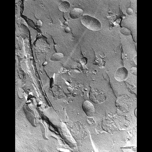
Freeze fracture of Vorticella microstoma. This cell had been placed in a relaxing medium of saturated sodium pyrophosphate plus 0.01M EDTA (Stain Tech. 47:37, 1972) for 3 hours. A myoneme enclosed by ER membrane is fractured at the cell’s periphery. In this cell it appears that the ER wraps around the myoneme. The P-fracture face of the plasma membrane is studded with many IMPs of significant size in contrast to the E-face of the outer alveolar membrane which has few IMPs. Bits of the P-face of the inner alveolar membrane show many IMPs on that face also. The P-face of the cytosolic-touching membrane of the ER and the E-face of the myonemal-touching membrane of the ER are visible. Linear arrays of IMPs on the E-face next to the myoneme may be transmembrane proteins associated with the linkage complex. Mitochondria are the most prominent organelles in the cytosol and their tubular cristae sometimes show signs of decoration with F1F0 ATP synthases. large arrow, direction of shadowing of freeze fracture replica. TEM taken on 11/11/75 by R. Allen with JEM 100B operating at 80kV. Neg. 11,015X. Bar = 0.5µm. A print of the negative was scanned and processed in Photoshop. This image is best used for qualitative analysis. There is a high resolution version of this image in the library (CIL:36275) which is available for quantitative analysis. Additional information available at (http://www5.pbrc.hawaii.edu/allen/).
| Spatial Axis | Image Size | Pixel Size |
|---|---|---|
| X | 2942px | —— |
| Y | 3672px | —— |