Alternate header for print version


Licensing
Public Domain: This image is in the public domain and thus free of any copyright restrictions. However, as is the norm in scientific publishing and as a matter of courtesy, any user should credit the content provider for any public or private use of this image whenever possible. Learn more
tweet  
*CIL – Cell Image Library accession number. Please use this to reference an image.

CIL:36293*  Cite 
Description

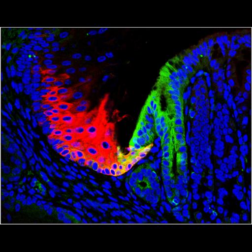
Embryonic cell precursors (green) of Barrett’s esophagus at the junction between the stomach and esophagus (red). Barrett's esophagus arrises not from mutant cells in the esophagus but rather a small group of previously overlooked cells in all adults that can rapidly expand to cancer precursors when the normal esophagus is damaged by acid. The red antibody is against keratin 6 expressed on junctional squamous cells, whereas the green antibody is to keratin 7 expressed in embryonic precursors of Barrett's that reside at the junction.

Biological Sources
NCBI Organism Classification
Mus musculus
Cell Type
squamous basal cells
juctional squamous cells
Biological Context
Biological Process
p63-/- mice
Human Disease
Barrett's esophagus
Attribution
Names
Xia Wang
Hong Ouyang
Yusuke Yamamoto
Pooja Ashok Kumar
Tay Seok Wei
Rania Dagher
Matthew Vincent
Xin Lu
Andrew M. Bellizzi
Khek Yu Ho
Christopher P. Crum
Wa Xian
Frank McKeon
Published
Cell. 2011 Jun 24;145(7):1023-35.
Pubmed
21703447
Link
Biomedical Beat
Focus - Harvard Medical School
ScienceDirect
Citation
Digital Object Identifier (DOI)
doi:10.7295/W9CIL36293
Archival Resource Key (ARK)
ark:/b7295/w9cil36293
Imaging
Image Type
recorded image
Image Mode
fluorescence microscopy
Parameters Imaged
fluorescence emission
Sample Preparation
Methods
chemically fixed tissue
Relation To Intact Cell
whole mounted tissue
Dimensions
Spatial Axis Image Size Pixel Size
X 3666px ——
Y 2850px ——