Alternate header for print version


Licensing
Public Domain: This image is in the public domain and thus free of any copyright restrictions. However, as is the norm in scientific publishing and as a matter of courtesy, any user should credit the content provider for any public or private use of this image whenever possible. Learn more
tweet  
*CIL – Cell Image Library accession number. Please use this to reference an image.

CIL:36592*  Cite 
Description

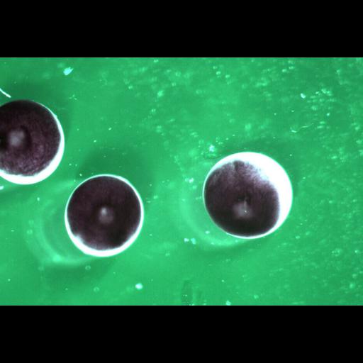
Time series of images of fertilized Xenopus laevis embryos with their vitellin membrane removed. Embryos were incubated plus/minus the actin depolymerizer cytochalsin B.

Technical Details

Images were collected with a dissecting microscope.

Biological Sources
NCBI Organism Classification
Xenopus laevis
Cell Type
fertilized embryo
Cellular Component
cell
Biological Context
Biological Process
cytochalasin D tratment
Xenopus Anatomy
embryo
Attribution
Names
Martin Wuehr
Timothy Mitchison
Citation
Digital Object Identifier (DOI)
doi:10.7295/W9CIL36592
Archival Resource Key (ARK)
ark:/b7295/w9cil36592
Grouping This image is part of a group.
Imaging
Image Type
recorded image
Image Mode
bright-field microscopy
Parameters Imaged
absorption of illumination
Source of Contrast
differences in intrinsic optical density
Visualization Methods
visualization of contiguous regions
Processing History
unprocessed raw data
Sample Preparation
Methods
living tissue
Relation To Intact Cell
whole mounted tissue
Dimensions
Spatial Axis Image Size Pixel Size
X 936px ——
Y 641px ——