Alternate header for print version


Licensing
Public Domain: This image is in the public domain and thus free of any copyright restrictions. However, as is the norm in scientific publishing and as a matter of courtesy, any user should credit the content provider for any public or private use of this image whenever possible. Learn more
tweet  
*CIL – Cell Image Library accession number. Please use this to reference an image.

CIL:36601*  Cite 
Description

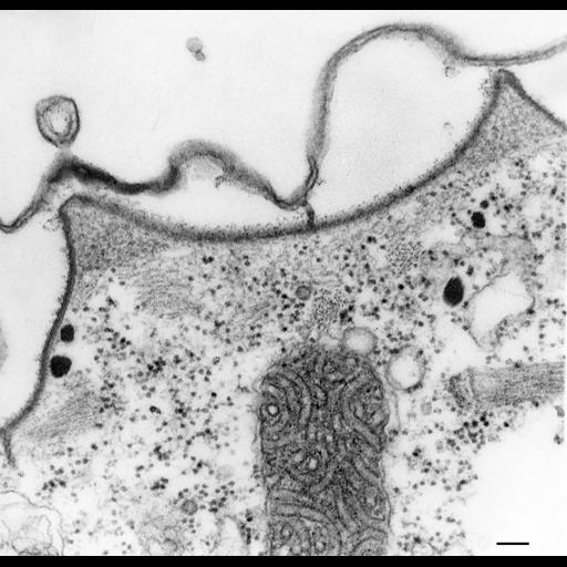
This view shows a transverse ridge cut with its sloping side lying tangentially in the section plane. The granulo-fibrillar filled peaks are prominent. Striated bands appear sectioned closer to their mid-sections, probably in areas of filament overlap. The outer alveolar membrane is not usually continuous with the inner alveolar membrane at the tips of the ridges. TEM taken on 11/11/70 by R. Allen with Hitachi HU11A operating at 60kV. Neg. 37,700X. Bar = 0.1µm.

Technical Details

Standard glutaraldehyde fixation followed by osmium tetroxide, dehydrated in alcohol and embedded in an epoxy resin. Microtome sections prepared at approximately 75nm thickness. The negative was printed to paper and the image was scanned to Photoshop. This digitized image is available for qualitative analysis. There is a high resolution version of this image in the library (CIL:39311) which is available for quantitative analysis. Additional information available at (http://www5.pbrc.hawaii.edu/allen/).

Biological Sources
NCBI Organism Classification
Paramecium multimicronucleatum
Cell Type
cell by organism
eukaryotic cell
Eukaryotic Protist
Ciliated Protist
Cellular Component
pellicle
cell cortex
Attribution
Names
Richard Allen (University of Hawaii)
Citation
Digital Object Identifier (DOI)
doi:10.7295/W9CIL36601
Archival Resource Key (ARK)
ark:/b7295/w9cil36601
Grouping This image is part of a group.
Imaging
Image Type
transmission electron microscopy (TEM)
illumination by electrons
Image Mode
detection of electrons
film
Parameters Imaged
electron density
Source of Contrast
stain with broad specificity
Visualization Methods
stain with broad specificity
uranyl salt
lead salt
Processing History
recorded image
film
Print from negative scanned for Photoshop.
Data Qualifiers
processed data
Dimensions
Spatial Axis Image Size Pixel Size
X 2368px ——
Y 2283px ——