Alternate header for print version


Licensing
Public Domain: This image is in the public domain and thus free of any copyright restrictions. However, as is the norm in scientific publishing and as a matter of courtesy, any user should credit the content provider for any public or private use of this image whenever possible. Learn more
tweet  
*CIL – Cell Image Library accession number. Please use this to reference an image.

CIL:36670*  Cite 
Description

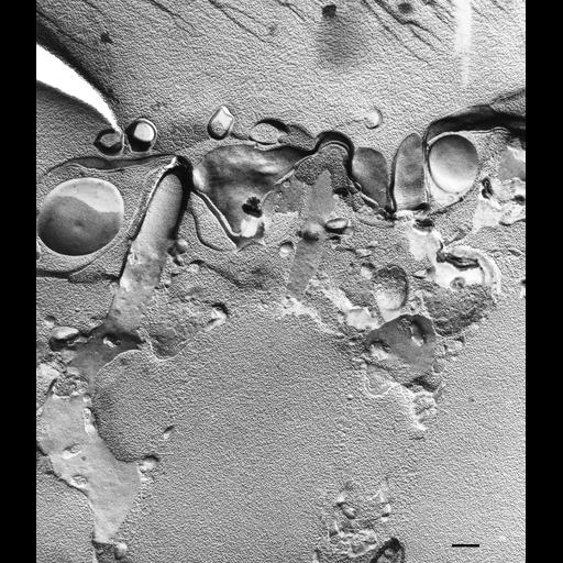
In a conventional freeze fracture view the membrane around the trichocyst tip is decorated with striations and IMPs. These decorations are found at the site where the trichocyst tip is associated with the alveolar membrane in the septum. Pores in the septum connect alveoli and organized IMPs at the base of the cilium indicate an association of the ciliary membrane with the proximal ends of the 9 doublets of the cilium and with the transition zone between the basal body and cilium. TEM taken on 9/27/78 by R. Allen with Hitachi HU11A operating at 75kV. Neg. 14,750X. Bar = 0.25µm.

Biological Sources
NCBI Organism Classification
Paramecium multimicronucleatum
Cell Type
cell by organism
eukaryotic cell
Eukaryotic Protist
Ciliated Protist
Cellular Component
plasma membrane
trichocyst
Biological Context
Biological Process
plasma membrane organization
Attribution
Names
Richard Allen (University of Hawaii)
Citation
Digital Object Identifier (DOI)
doi:10.7295/W9CIL36670
Archival Resource Key (ARK)
ark:/b7295/w9cil36670
Imaging
Image Type
transmission electron microscopy (TEM)
illumination by electrons
Image Mode
electron density
Parameters Imaged
elastic scattering of electrons
electron density
elevation
Source of Contrast
differences in deposition of metal shadow
Visualization Methods
transmission electron microscopy (TEM)
Processing History
freeze_fracture/freeze_etch
shadowing and plating
recorded image
film
Print of negative scanned for Photoshop
Data Qualifiers
processed data
Dimensions
Spatial Axis Image Size Pixel Size
X 2196px ——
Y 2526px ——