Alternate header for print version


Licensing
Public Domain: This image is in the public domain and thus free of any copyright restrictions. However, as is the norm in scientific publishing and as a matter of courtesy, any user should credit the content provider for any public or private use of this image whenever possible. Learn more
tweet  
*CIL – Cell Image Library accession number. Please use this to reference an image.

CIL:36712*  Cite 
Description

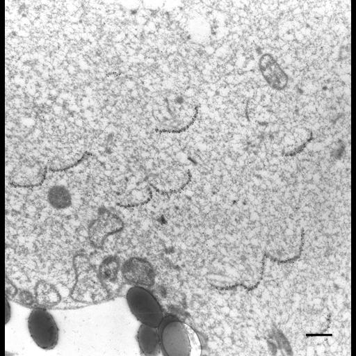
The cytopharyngeal ribbons of microtubules tend to curve into a “C” shape away from the cytopharynx but they still remain connected as a ribbon and are heavily labeled by immunogold when exposed to anti-b-tubulin. TEM taken on 2/27/94 by R. Allen with Zeiss 10A operating at 80kV. Neg. 9,780X. Bar = 0.5µm.

Biological Sources
NCBI Organism Classification
Paramecium multimicronucleatum
Cell Type
cell by organism
eukaryotic cell
Eukaryotic Protist
Ciliated Protist
Cellular Component
microtubule associated complex
oral apparatus
Attribution
Names
Richard Allen (University of Hawaii)
Citation
Digital Object Identifier (DOI)
doi:10.7295/W9CIL36712
Archival Resource Key (ARK)
ark:/b7295/w9cil36712
Imaging
Image Type
transmission electron microscopy (TEM)
illumination by electrons
Image Mode
detection of electrons
film
Parameters Imaged
electron density
Source of Contrast
intrinsic mass distribution
stain with broad specificity
Visualization Methods
stain with broad specificity
uranyl salt
gold
Processing History
recorded image
film
Print from negative scanned for Photoshop.
Data Qualifiers
processed data
Sample Preparation
Methods
glutaraldehyde fixed tissue
tissue in vitreous ice embedment
microtome-sectioned tissue
monoclonal antibody
Relation To Intact Cell
microtome-sectioned tissue
Dimensions
Spatial Axis Image Size Pixel Size
X 2160px ——
Y 2220px ——