Alternate header for print version


Licensing
Public Domain: This image is in the public domain and thus free of any copyright restrictions. However, as is the norm in scientific publishing and as a matter of courtesy, any user should credit the content provider for any public or private use of this image whenever possible. Learn more
tweet  
*CIL – Cell Image Library accession number. Please use this to reference an image.

CIL:36739*  Cite 
Description

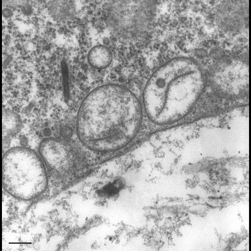
Secondary lysosomes dock only with phagoacidosome membrane at a distance of 40nm. As during acidosome fusion with phagosomes, fusion of the many lysosomes with the phagoacidosome occurs at nearly the same time. Some small vesicles can be found close to the lysosomal surface. Such small vesicles might represent primary lysosomes originating from Golgi complexes, such as the coated vesicle in the upper left corner of this micrograph might suggest. TEM taken on 8/16/80 by R. Allen with Hitachi HU11A operating at 75kV. Neg. 31,500X. Bar = 0.2µm.

Technical Details

Standard glutaraldehyde fixation followed by osmium tetroxide, dehydrated in alcohol and embedded in an epoxy resin. Microtome sections prepared at approximately 75nm thickness. The negative was printed to paper and the image was scanned to Photoshop. This digitized image is available for qualitative analysis. Additional information available at (http://www5.pbrc.hawaii.edu/allen/).

Biological Sources
NCBI Organism Classification
Paramecium multimicronucleatum
Cell Type
cell by organism
eukaryotic cell
Eukaryotic Protist
Ciliated Protist
Cellular Component
phagosome
food vacuole
primary lysosome
Biological Context
Biological Process
digestive system process
Attribution
Names
Richard Allen (University of Hawaii)
Citation
Digital Object Identifier (DOI)
doi:10.7295/W9CIL36739
Archival Resource Key (ARK)
ark:/b7295/w9cil36739
Imaging
Image Type
transmission electron microscopy (TEM)
illumination by electrons
Image Mode
detection of electrons
film
Parameters Imaged
electron density
Source of Contrast
stain with broad specificity
Visualization Methods
stain with broad specificity
osmium tetroxide
uranyl salt
lead salt
Processing History
recorded image
film
Print from negative scanned for Photoshop.
Data Qualifiers
processed data
Dimensions
Spatial Axis Image Size Pixel Size
X 2280px ——
Y 2316px ——