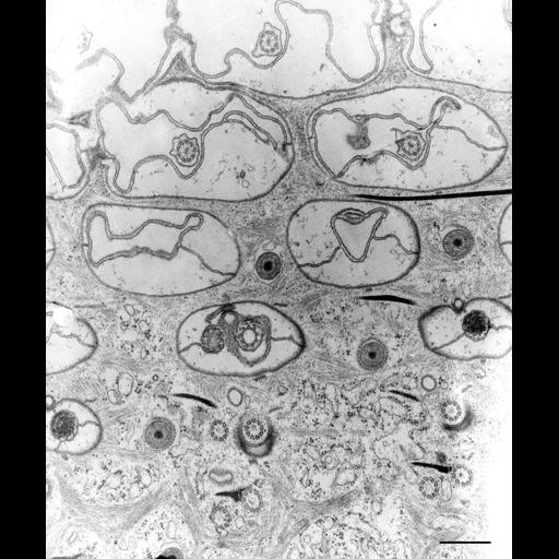
A grazing section through the cell’s surface shows segments of six rows of cilia running across the figure sectioned superfically at the top and deeper into the cell as one progresses down the figure. Crests of the polygonal ridges appear at the top of the figure where the expanded alveoli fill most of the space in the surface depressions. Septa, the borders of adjoining alveolar sacs, run between basal body/cilium complexes along a kinety. Trichocyst tips penetrate into the lateral ridges that separate the concave depressions of a kinety. Within the cortical cytosol a more superficial system of striated bands connects the slopes of adjacent ridges, laterally, longitudinally and diagonally. Internal to the striated bands is a meshwork of centrin-containing fibrous bundles called the infraciliary lattice. This meshwork surrounds the proximal half of basal bodies but does not insert into the pellicular membranes. Collidine-buffered fixative. TEM taken on 7/8/69 by R. Allen with Philips 300 operating at 60kV. Neg. 14,800X. Bar = 0.5µm.
Standard glutaraldehyde fixation followed by osmium tetroxide, dehydrated in alcohol and embedded in an epoxy resin. Microtome sections prepared at approximately 75nm thickness. The negative was printed to paper and the image was scanned to Photoshop. This digitized image is available for qualitative analysis. There is a high resolution version of this image in the library (CIL:38892) which is available for quantitative analysis. Additional information available at (http://www5.pbrc.hawaii.edu/allen/).
| Spatial Axis | Image Size | Pixel Size |
|---|---|---|
| X | 2856px | —— |
| Y | 3424px | —— |