Alternate header for print version


Licensing
Attribution Non-Commercial Share Alike:This image is licensed under a Creative Commons Attribution, Non-Commercial Share Alike License. View License Deed | View Legal Code
tweet  
*CIL – Cell Image Library accession number. Please use this to reference an image.

CIL:36760*  Cite 
Description

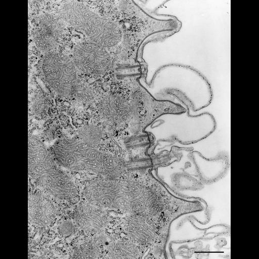
Septa of bordering alveoli when lying in the plane of section exhibit pores at their outer margins closest to the plasma membrane. This shows that there is physical continuity between the lumens of alveoli. Basal bodies and fibrous systems are found in the cellular cortex beneath the ridges. Cacodylate buffered fixative. TEM taken on 11/2/68 by R. Allen with Philips 300 operating at 60kV. Neg. 20,500X. Bar = 0.5µm. Published in J. Cell Biol. 49:1-20, 1971. Adapted with permission.

Technical Details

Standard glutaraldehyde fixation followed by osmium tetroxide, dehydrated in alcohol and embedded in an epoxy resin. Microtome sections prepared at approximately 75nm thickness. The negative was printed to paper and the image was scanned to Photoshop. This digitized image is available for qualitative analysis. There is a high resolution version of this image in the library (CIL:38896) which is available for quantitative analysis. Additional information available at (http://www5.pbrc.hawaii.edu/allen/).

Biological Sources
NCBI Organism Classification
Paramecium caudatum
Cell Type
cell by organism
eukaryotic cell
Eukaryotic Protist
Ciliated Protist
Cellular Component
pellicle
plasma membrane
cell cortex
Biological Context
Biological Process
plasma membrane organization
Attribution
Names
Richard Allen (University of Hawaii)
Published
J Cell Biol. 1971 Apr;49(1):1-20.
Pubmed
4102004
Citation
Digital Object Identifier (DOI)
doi:10.7295/W9CIL36760
Archival Resource Key (ARK)
ark:/b7295/w9cil36760
Grouping This image is part of a group.
Imaging
Image Type
transmission electron microscopy (TEM)
illumination by electrons
Image Mode
detection of electrons
film
Parameters Imaged
electron density
Source of Contrast
stain with broad specificity
Visualization Methods
stain with broad specificity
uranyl salt
osmium tetroxide
lead salt
Processing History
recorded image
film
Print from negative scanned for Photoshop.
Data Qualifiers
processed data
Dimensions
Spatial Axis Image Size Pixel Size
X 2148px ——
Y 2742px ——