Alternate header for print version


Licensing
Attribution Non-Commercial Share Alike:This image is licensed under a Creative Commons Attribution, Non-Commercial Share Alike License. View License Deed | View Legal Code
tweet  
*CIL – Cell Image Library accession number. Please use this to reference an image.

CIL:37186*  Cite 
Description

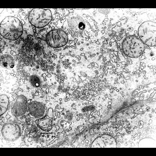
Transmission electron micrograph of thin section of rat liver with prominent Golgi and smooth ER associated vesicles. At lower right, the junction between hepatocytes harbors a bile canaliculus. The image illustrates the ethanol-stimulated accumulation of low density lipoprotein (LDL) in Golgi of the liver in rats fed ethanol (equivalent to 1 quart of single malt whisky) by gastric tube. This observation was the impetus for the strategy of purifying Golgi factions by allowing them to rise the surface of dense sucrose columns. Image made available by James D. Jamieson and the Department of Cell Biology, Yale University School of Medicine.

Technical Details

Original 3.25 in. x 4 in. lantern slides were scanned at 600dpi. Original Magnification: x19,000.

Biological Sources
NCBI Organism Classification
Rattus
Cell Type
hepatocyte
Cellular Component
Golgi apparatus
mitochondrion
cell-cell junction
low-density lipoprotein particle
smooth endoplasmic reticulum
Biological Context
Biological Process
LDL accumulation
Attribution
Names
George E. Palade
Link
George E. Palade EM Slide Collection
OTHER
02/01/1969
Citation
Digital Object Identifier (DOI)
doi:10.7295/W9CIL37186
Archival Resource Key (ARK)
ark:/b7295/w9cil37186
Grouping This image is part of a group.
Imaging
Image Type
recorded image
Image Mode
transmission electron microscopy (TEM)
Parameters Imaged
electron density
Source of Contrast
differences in adsorption or binding of stain
Visualization Methods
osmium tetroxide
Processing History
unprocessed raw data
Sample Preparation
Methods
chemically fixed tissue
Relation To Intact Cell
sectioned tissue
Dimensions
Spatial Axis Image Size Pixel Size
X 6000px ——
Y 5377px ——