Alternate header for print version


Licensing
Attribution Non-Commercial; No Derivatives:This image is licensed under a Creative Commons Attribution, Non-Commercial, No Derivatives License. View License Deed | View Legal Code
tweet  
*CIL – Cell Image Library accession number. Please use this to reference an image.

CIL:38809*  Cite 
Description

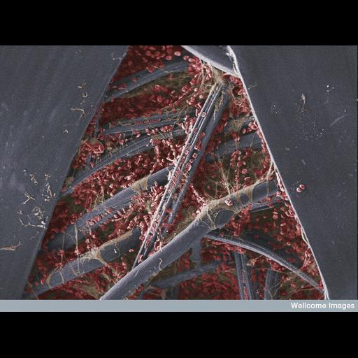
Scanning electron micrograph of a blood clot on the underside of a sticking plaster that has been used to treat a razor blade cut. Red blood cells (shown in red) and thin fibres of the protein fibrin (beige) can be seen between the gauze fibres of the plaster (blue-grey). Fibrin is a protein formed from the conversion of clotting factors in the blood; the fibrin fibers trap blood cells and platelets to form a solid clot. This not only prevents further bleeding but also protects the open wound from infection.

Technical Details

B0007385. 2011 Wellcome Image Award winner Scanning electron micrograph Collection: Wellcome Images Copyrighted work available under Creative Commons by-nc-nd 2.0 UK: England & Wales, see http://creativecommons.org/licenses/by-nc-nd/2.0/uk/

Biological Sources
NCBI Organism Classification
Homo sapiens
Cell Type
blood cell
Cellular Component
extracellular matrix part
cell surface
Biological Context
Biological Process
blood coagulation, fibrin clot formation
Attribution
Names
Anne Weston
Link
Wellcome Images
OTHER
LRI, CRUK
Citation
Digital Object Identifier (DOI)
doi:10.7295/W9CIL38809
Archival Resource Key (ARK)
ark:/b7295/w9cil38809
Imaging
Image Type
recorded image
Image Mode
scanning electron microscopy (SEM)
Parameters Imaged
elastic scattering of electrons
Source of Contrast
differences in deposition of metal shadow
Visualization Methods
visualization of contiguous regions
Processing History
colorized
Sample Preparation
Methods
chemically fixed tissue
Relation To Intact Cell
whole mounted tissue
Dimensions
Spatial Axis Image Size Pixel Size
X 774px ——
Y 576px ——