Alternate header for print version


Licensing
Public Domain: This image is in the public domain and thus free of any copyright restrictions. However, as is the norm in scientific publishing and as a matter of courtesy, any user should credit the content provider for any public or private use of this image whenever possible. Learn more
tweet  
*CIL – Cell Image Library accession number. Please use this to reference an image.

CIL:38861*  Cite 
Description

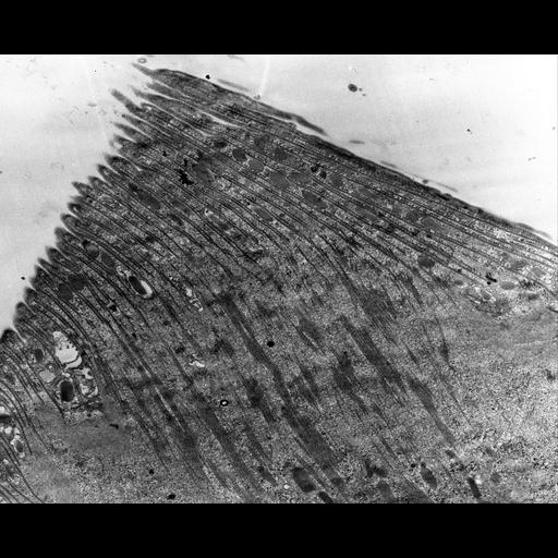
A high resolution view of the cytopharynx but in a dividing Didinium. This cytopharynx is in the proter (anterior) daughter cell. The lamellae, each consisting of a set of perpendicularly arranged microtubules cover this food-vacuole forming region. Vesicles lie near the cytopharyngeal membrane and fuse with the membrane. TEM taken on 5/20/69 by R. Allen with Philips 300 operating at 60kV. Neg. 6,370X.

Technical Details

The raw film was scanned with a Nikon Coolscan 9000ED. This digitized image is available for quantitative analysis. Standard glutaraldehyde fixation followed by osmium tetroxide, dehydrated in alcohol and embedded in an epoxy resin. Microtome sections prepared at approximately 75nm thickness. Additional information available at (http://www5.pbrc.hawaii.edu/allen/).

Biological Sources
NCBI Organism Classification
Didinium nasutum
Cell Type
cell by organism
eukaryotic cell
Eukaryotic Protist
Ciliated Protist
Cell Line
Carolina Biological Supply Company, NC, U.S.A.
Cellular Component
cell cortex
cortical microtubule cytoskeleton
Attribution
Names
Richard Allen
Citation
Digital Object Identifier (DOI)
doi:10.7295/W9CIL38861
Archival Resource Key (ARK)
ark:/b7295/w9cil38861
Grouping This image is part of a group.
Imaging
Image Type
transmission electron microscopy (TEM)
illumination by electrons
Image Mode
detection of electrons
film
Parameters Imaged
electron density
Source of Contrast
stain with broad specificity
Visualization Methods
stain with broad specificity
osmium tetroxide
uranyl salt
lead salt
Processing History
recorded image
film
unprocessed raw data
Data Qualifiers
raw, unprocessed data
suitable for spatial measurements
Dimensions
Spatial Axis Image Size Pixel Size
X 4000px 1.6nm
Y 3224px 1.6nm