Alternate header for print version


Licensing
Public Domain: This image is in the public domain and thus free of any copyright restrictions. However, as is the norm in scientific publishing and as a matter of courtesy, any user should credit the content provider for any public or private use of this image whenever possible. Learn more
tweet  
*CIL – Cell Image Library accession number. Please use this to reference an image.

CIL:38898*  Cite 
Description

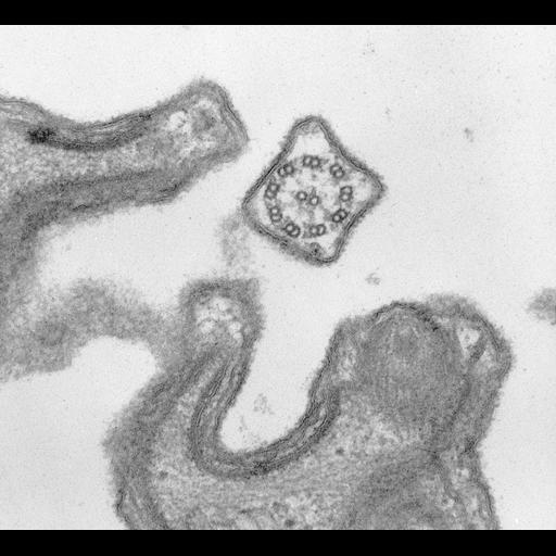
Cross section of a cilium showing the 9 + 2 axoneme. Two dynein arms, inner and outer, of heterogeneous appearance arise from the complete microtubule of each doublet. Spokes extend from the doublets toward the central pair. Other densities extend from the central pair and between some of the outer doublets and the membrane. A heavy glycocalyx (polysaccharide coat) is found on the outside of the ciliary membrane as well as the plasma membrane. TEM taken on 2/3/69 by R. Allen with Philips 300 operating at 60kV. Neg. 47,100X.

Technical Details

Standard glutaraldehyde fixation followed by osmium tetroxide, dehydrated in alcohol and embedded in an epoxy resin. Microtome sections prepared at approximately 75nm thickness. The raw negative was scanned with an Epson Perfection V750 Pro and this high resolution image is best used for quantitative analysis. Additional information available at (http://www5.pbrc.hawaii.edu/allen/).

Biological Sources
NCBI Organism Classification
Paramecium caudatum
Cell Type
cell by organism
eukaryotic cell
Eukaryotic Protist
Ciliated Protist
Cellular Component
cilium
glycocalyx
Biological Context
Biological Process
ciliary or flagellar motility
plasma membrane organization
Attribution
Names
Richard Allen (University of Hawaii)
Citation
Digital Object Identifier (DOI)
doi:10.7295/W9CIL38898
Archival Resource Key (ARK)
ark:/b7295/w9cil38898
Grouping This image is part of a group.
Imaging
Image Type
transmission electron microscopy (TEM)
illumination by electrons
Image Mode
detection of electrons
film
Parameters Imaged
electron density
Source of Contrast
stain with broad specificity
Visualization Methods
stain with broad specificity
uranyl salt
lead salt
osmium tetroxide
Processing History
recorded image
film
unprocessed raw data
Data Qualifiers
raw, unprocessed data
suitable for spatial measurements
Dimensions
Spatial Axis Image Size Pixel Size
X 3472px 0.42nm
Y 3156px 0.42nm