Alternate header for print version


Licensing
Attribution Non-Commercial; No Derivatives:This image is licensed under a Creative Commons Attribution, Non-Commercial, No Derivatives License. View License Deed | View Legal Code
tweet  
*CIL – Cell Image Library accession number. Please use this to reference an image.

CIL:38933*  Cite 
Description

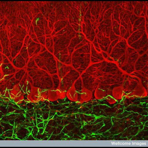
This confocal micrograph shows specialized cells named Purkinje cells (red) that are found in a part of the brain called the cerebellum. They send out vast numbers of branches that make connections with other cells in the cerebellum. This part of the brain co-ordinates your voluntary movements and keeps you oriented in space. It also plays a part in learning physical skills - like riding a bike or playing the piano.

Technical Details

B0006224 Purkinje cells in the cerebellum. Wellcome Images available under the following creative commons usage http://creativecommons.org/licenses/by-nc-nd/2.0/uk/

Biological Sources
Cell Type
Purkinje cell
Cellular Component
neuron projection
neuronal cell body
Biological Context
Biological Process
cerebellar Purkinje cell layer morphogenesis
Attribution
Names
Ludovic Colin
Link
Wellcome Images
Citation
Digital Object Identifier (DOI)
doi:10.7295/W9CIL38933
Archival Resource Key (ARK)
ark:/b7295/w9cil38933
Imaging
Image Type
recorded image
Image Mode
confocal microscopy
Parameters Imaged
fluorescence emission
Source of Contrast
compartmentalization of stain or label
Processing History
unprocessed raw data
Sample Preparation
Relation To Intact Cell
sectioned tissue
Dimensions
Spatial Axis Image Size Pixel Size
X 584px ——
Y 576px ——