Alternate header for print version


Licensing
Attribution Non-Commercial; No Derivatives:This image is licensed under a Creative Commons Attribution, Non-Commercial, No Derivatives License. View License Deed | View Legal Code
tweet  
*CIL – Cell Image Library accession number. Please use this to reference an image.

CIL:38972*  Cite 
Description

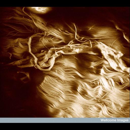
Confocal image showing damaged collagen fibres in a ruptured tendon. The area of wavey fibres to the lower right shows the normal healthy appearence of tendon. The waviness allows the whole tendon to have a small amount of elasticity (2-10%) as the collagen fibres themselves do not stretch. The field of view is approximately 220 microns across

Technical Details

Wellcome Images available under the following creative commons usage http://creativecommons.org/licenses/by-nc-nd/2.0/uk/

Biological Sources
Cell Type
tendon cell
Cellular Component
fibrillar collagen
Biological Context
Biological Process
tendon rupture
Attribution
Names
Martin Knight
Link
Wellcome Images
Citation
Digital Object Identifier (DOI)
doi:10.7295/W9CIL38972
Archival Resource Key (ARK)
ark:/b7295/w9cil38972
Imaging
Image Type
recorded image
Image Mode
confocal microscopy
Visualization Methods
visualization of contiguous regions
Sample Preparation
Methods
chemically fixed tissue
Relation To Intact Cell
whole mounted tissue
Dimensions
Spatial Axis Image Size Pixel Size
X 644px ——
Y 576px ——