Alternate header for print version


Licensing
Attribution Non-Commercial; No Derivatives:This image is licensed under a Creative Commons Attribution, Non-Commercial, No Derivatives License. View License Deed | View Legal Code
tweet  
*CIL – Cell Image Library accession number. Please use this to reference an image.

CIL:38976*  Cite 
Description

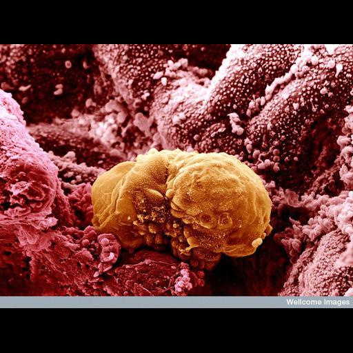
Scanning electron micrograph of a 6 day old human embryo beginning to implant into the lining of the uterus (endometrium). As implantation progresses, the inner cell mass begins to form into the bilaminar disc. The two layers are called the epiblast and the hypoblast. An embryo that has been in culture for up to 14 days will remain at this stage of development. Such cultured embryos remain alive but do not progress as they would in the womb.

Technical Details

B0003308 6 day old human embryo implanting. Wellcome Images available under the following creative commons usage http://creativecommons.org/licenses/by-nc-nd/2.0/uk/

Biological Sources
NCBI Organism Classification
Homo sapiens
Cell Type
embryonic gland hemocyte
Cellular Component
cell surface
Biological Context
Biological Process
embryo implantation
Human Development Anatomy
endometrium
bilaminar disc
epiblast
hypoblast
Attribution
Names
Yorgos Nikas
Link
Wellcome Images
Citation
Digital Object Identifier (DOI)
doi:10.7295/W9CIL38976
Archival Resource Key (ARK)
ark:/b7295/w9cil38976
Imaging
Image Type
recorded image
Image Mode
scanning electron microscopy (SEM)
Parameters Imaged
elastic scattering of electrons
Source of Contrast
differences in deposition of metal shadow
Visualization Methods
visualization of contiguous regions
Processing History
colorized
Sample Preparation
Methods
chemically fixed tissue
Relation To Intact Cell
whole mounted tissue
Dimensions
Spatial Axis Image Size Pixel Size
X 770px ——
Y 576px ——