Alternate header for print version


Licensing
Attribution Non-Commercial; No Derivatives:This image is licensed under a Creative Commons Attribution, Non-Commercial, No Derivatives License. View License Deed | View Legal Code
tweet  
*CIL – Cell Image Library accession number. Please use this to reference an image.

CIL:39020*  Cite 
Description

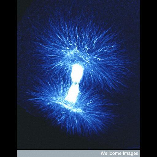
Fluorescent micrograph showing the mitotic spindle, which is composed of microtubules. During cell division the chromosomes duplicate and are pulled to opposite poles by the spindle, once the chromosomes have separated the cell divides and forms two new daughter cells.

Technical Details

B0007804 Mitotic spindle. Wellcome Images available under the following creative commons usage http://creativecommons.org/licenses/by-nc-nd/2.0/uk/

Biological Sources
Cellular Component
spindle
Biological Context
Biological Process
mitosis
mitotic anaphase
Attribution
Names
B. Strauss
Link
Wellcome Images
Citation
Digital Object Identifier (DOI)
doi:10.7295/W9CIL39020
Archival Resource Key (ARK)
ark:/b7295/w9cil39020
Imaging
Image Type
recorded image
Image Mode
confocal microscopy
Parameters Imaged
fluorescence emission
Source of Contrast
distribution of a specific protein
Processing History
unprocessed raw data
Sample Preparation
Relation To Intact Cell
dispersed cells in vitro
Dimensions
Spatial Axis Image Size Pixel Size
X 453px ——
Y 576px ——