Alternate header for print version


Licensing
Attribution Non-Commercial; No Derivatives:This image is licensed under a Creative Commons Attribution, Non-Commercial, No Derivatives License. View License Deed | View Legal Code
tweet  
*CIL – Cell Image Library accession number. Please use this to reference an image.

CIL:39051*  Cite 
Description

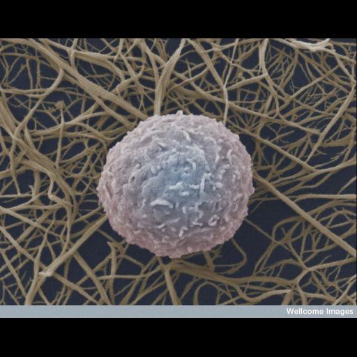
Colorized scanning electron micrograph (SEM) of a human white blood cell (leukocyte) on a mesh of fibrin. Fibrin is an important protein in the formation of blood clots.

Technical Details

B0007654 Human white blood cell. Wellcome Images available under the following creative commons usage http://creativecommons.org/licenses/by-nc-nd/2.0/uk/

Biological Sources
NCBI Organism Classification
Homo sapiens
Cell Type
blood cell
Cellular Component
extracellular matrix
cell surface
Biological Context
Biological Process
blood coagulation, fibrin clot formation
Attribution
Names
Anne Weston
Link
Wellcome Images
OTHER
LRI, CRUK
Citation
Digital Object Identifier (DOI)
doi:10.7295/W9CIL39051
Archival Resource Key (ARK)
ark:/b7295/w9cil39051
Imaging
Image Type
recorded image
Image Mode
scanning electron microscopy (SEM)
Parameters Imaged
elastic scattering of electrons
Source of Contrast
differences in deposition of metal shadow
Visualization Methods
visualization of contiguous regions
Processing History
colorized
Sample Preparation
Methods
chemically fixed tissue
Dimensions
Spatial Axis Image Size Pixel Size
X 735px ——
Y 576px ——