Alternate header for print version


Licensing
Attribution Non-Commercial Share Alike:This image is licensed under a Creative Commons Attribution, Non-Commercial Share Alike License. View License Deed | View Legal Code
tweet  
*CIL – Cell Image Library accession number. Please use this to reference an image.

CIL:39334*  Cite 
Description

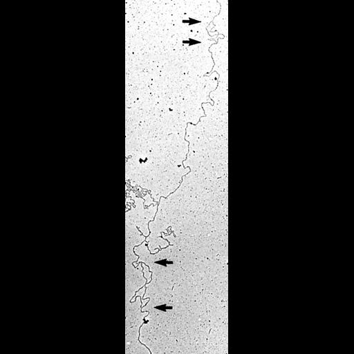
Nuclear DNA was prepared from synchronously replicating nuclei of Physarum polycephalum, mixed with cytochrome c and formamide, and spread on the surface of a hypophase consisting of 10 mM Tris, 1 mM EDTA and 10% formamide. The surface containing the DNA-cytochrome c complexes was picked up on carbon-formvar grids, stained with ethanolic phosphotungstic acid, and rotary shadowed with Pt/Pd prior to TEM examination. Arrows point to replication forks.

Technical Details

See Fig 3A in: F Haugli et al. 1982 DNA replication in Physarum polycephalum: electron microscopic analysis patterns of DNA replication in the presence of cycloheximide. J Cell Biol 95:323-331.

Biological Sources
NCBI Organism Classification
Physarum polycephalum
Cell Type
plasmodium
Cell Line
TU291
Cellular Component
DNA
Biological Context
Biological Process
DNA replication
DNA metabolic process
Attribution
Names
Finn Haugli
Rolf Andreassen
Steiner Funderud
Published
F Haugli et al. 1982 DNA replication in Physarum polycephalum: electron microscopic analysis patterns of DNA replication in the presence of cycloheximide. J Cell Biol 95:323-331.
Pubmed
7142290
Citation
Digital Object Identifier (DOI)
doi:10.7295/W9CIL39334
Archival Resource Key (ARK)
ark:/b7295/w9cil39334
Imaging
Image Type
film
Image Mode
transmission electron microscopy (TEM)
Parameters Imaged
electron density
Source of Contrast
differences in deposition of metal shadow
Visualization Methods
shadowing and plating
tungsten compound
Processing History
montage
Data Qualifiers
processed data
Sample Preparation
Relation To Intact Cell
isolated subcellular component
Dimensions
Spatial Axis Image Size Pixel Size
X 420px ——
Y 1443px ——