Alternate header for print version


Licensing
Public Domain: This image is in the public domain and thus free of any copyright restrictions. However, as is the norm in scientific publishing and as a matter of courtesy, any user should credit the content provider for any public or private use of this image whenever possible. Learn more
tweet  
*CIL – Cell Image Library accession number. Please use this to reference an image.

CIL:39341*  Cite 
Description

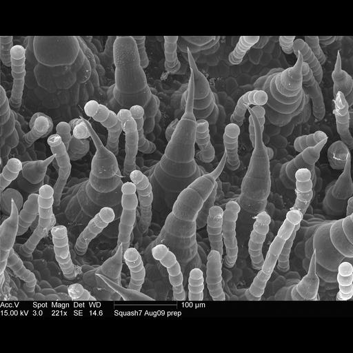
Scanning electron microscope image of sunflower lower leaf surface. This is part of an image series containing images CIL:39341-39343, and 39345.

Technical Details

Instrument: FEI XL-30 FEG ESEM. Technical protocol available at http://remf.dartmouth.edu:8080/EM-Wiki/36

Biological Sources
NCBI Organism Classification
Helianthus annuus
Attribution
Names
Louisa Howard
Link
Dartmouth College EM Facility
Citation
Digital Object Identifier (DOI)
doi:10.7295/W9CIL39341
Archival Resource Key (ARK)
ark:/b7295/w9cil39341
Grouping This image is part of a group.
Imaging
Image Type
recorded image
Image Mode
scanning electron microscopy (SEM)
Parameters Imaged
elastic scattering of electrons
Source of Contrast
differences in deposition of metal shadow
Visualization Methods
uranyl salt
shadowing and plating
Processing History
unprocessed raw data
Sample Preparation
Methods
glutaraldehyde fixed tissue
formaldehyde fixed tissue
Relation To Intact Cell
whole mounted tissue
Dimensions
Spatial Axis Image Size Pixel Size
X 2576px ——
Y 2056px ——