Alternate header for print version


Licensing
Attribution Non-Commercial Share Alike:This image is licensed under a Creative Commons Attribution, Non-Commercial Share Alike License. View License Deed | View Legal Code
tweet  
*CIL – Cell Image Library accession number. Please use this to reference an image.

CIL:39736*  Cite 
Description

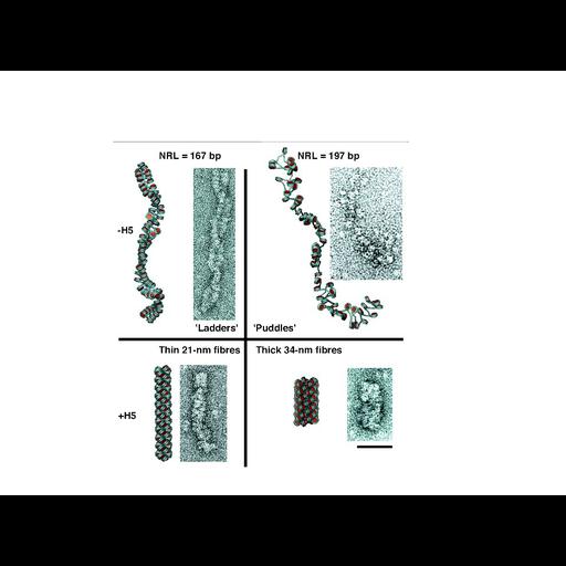
Images of negatively stained chromatin fibers prepared from tandem arrays of a nucleosome positioning sequence reconstituted with purified histones together with the results of Monte Carlo simulations of chromatin folding. Top left - 167 bp repeat with no linker histone; upper right - 197 bp with without linker histone; lower left 167 bp with linker histone H5; lower right 197 bp with linker histone. . See: Fig 4 in Routh et al. (2008) Nucleosome repeat length and linker histonen stoichiometry determine chromatin fiber structure. Proc Natl Acad Sci USA 105:8872-8877.

Technical Details

Reconstituted chromatin was fixed with 0.1% glutaraldehyde for 30 min, deposited on glow discharged carbon films, negatively stained with aqueous uranyl acetate, and examined with a FEI Tecnai F30 TEM operated at 200 KV. See also: Routh et al. (2008) Nucleosome repeat length and linker histonen stoichiometry determine chromatin fiber structure. Proc Natl Acad Sci USA 105:8872-8877.

Biological Sources
Cellular Component
nuclear chromatin
chromatin
Attribution
Names
Andrew Routh
Sara Sandin
Daniela Rhodes
Published
A Routh et al. (2008) Nucleosome repeat length and linker histonen stoichiometry determine chromatin fiber structure. Proc Natl Acad Sci USA 105:8872-8877
Pubmed
18583476
Link
Proceedings National Academy of Sciences USA
Citation
Digital Object Identifier (DOI)
doi:10.7295/W9CIL39736
Archival Resource Key (ARK)
ark:/b7295/w9cil39736
Grouping This image is part of a group.
Imaging
Image Type
recorded image
portrayed image
Image Mode
transmission electron microscopy (TEM)
Parameters Imaged
electron density
Source of Contrast
differences in adsorption or binding of stain
Visualization Methods
negative staining
uranyl salt
Processing History
montage
Data Qualifiers
processed data
Sample Preparation
Methods
glutaraldehyde fixed tissue
Relation To Intact Cell
isolated subcellular component
Dimensions
Spatial Axis Image Size Pixel Size
X 960px ——
Y 720px ——