Alternate header for print version


Licensing
Public Domain: This image is in the public domain and thus free of any copyright restrictions. However, as is the norm in scientific publishing and as a matter of courtesy, any user should credit the content provider for any public or private use of this image whenever possible. Learn more
tweet  
*CIL – Cell Image Library accession number. Please use this to reference an image.

CIL:39746*  Cite 
Description

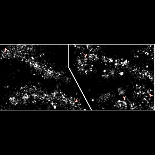
PALM images of two regions of Drosophila metaphase chromosomes with red arrowheads pointing to filamentous structures. Overview images of the chromosome clusters are seen the the grouped image (CIL:39747) See Fig 3 in: A Matsuda et al. (2010) Condensed mitotic chromosme structure at nanometer resolution using PALM and EGFP-histones. PLOSone 5:e12768.

Technical Details

PALM was achieved using a custom wide-field microscope platform with 100x NA 1.4 objective lens or 150X TIRF NA 1.45 lens, yielding a pixel size at the CCD camera of 0.0792 or 0.0528 micrometers respectively. See also: A Matsuda et al. (2010) Condensed mitotic chromosme structure at nanometer resolution using PALM and EGFP-histones. PLOSone 5:e12768.

Biological Sources
NCBI Organism Classification
Drosophila melanogaster
Cell Type
early embryonic cell
Cellular Component
nuclear chromosome
chromatin
Biological Context
Biological Process
DNA packaging
chromatin organization
Attribution
Names
A Matsuda
L Shao
J Boulanger
C Kervrann
PM Carlton
P Kner
D Agard
J Sedat
Published
A Matsuda et al. (2010) Condensed mitotic chromosme structure at nanometer resolution using PALM and EGFP-histones. PLOSone 5:e12768
Pubmed
20856676
Link
PLOSone
Citation
Digital Object Identifier (DOI)
doi:10.7295/W9CIL39746
Archival Resource Key (ARK)
ark:/b7295/w9cil39746
Grouping This image is part of a group.
Imaging
Image Type
portrayed image
Image Mode
PALM (photoactivation localization microscopy)
Parameters Imaged
fluorescence emission
Source of Contrast
distribution of a specific protein
Visualization Methods
EGFP
4',6-diamidino-2-phenylindole (DAPI)
Processing History
PALM reconstruction
Data Qualifiers
processed data
Sample Preparation
Methods
formaldehyde fixed tissue
Relation To Intact Cell
whole mounted tissue
Dimensions
Spatial Axis Image Size Pixel Size
X 942px ——
Y 403px ——