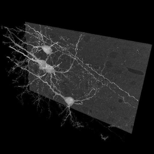
Maximum intensity projection of Golgi impregnated pyramidal neurons in the neocortex of an adult mouse, imaged via serial block-face scanning electron microscopy. Contrast is reversed so that Golgi stain appears bright against the unstained background. This image has been downsampled from the raw data image which can be accessed using the link provided to the Cell Centered Database.
Perfused with 4% PFA, 0.1% glut. Postfixed 1 hour in same fix on ice. Left hemisphere cut into 2 mm thick coronal slabs. Tissue placed into 2.3% potassium dichromate / 0.4% OsO4 for 3 days at room temp. Tissue blotted with filter paper and then placed in 0.7% silver nitrate in dark for 3 days. Tissue was dehydrated, infiltrated and embedded in Durcupan in Beem capsules. Serial section images were gathered on an FEI Quanta 200 FEG SEM with Gatan 3 View microscope, magnification, 600.0 X, accelerating voltage, 2.0 kV.
| Spatial Axis | Image Size | Pixel Size |
|---|---|---|
| X | 512px | —— |
| X | 435px | —— |