Alternate header for print version


Licensing
Public Domain: This image is in the public domain and thus free of any copyright restrictions. However, as is the norm in scientific publishing and as a matter of courtesy, any user should credit the content provider for any public or private use of this image whenever possible. Learn more
tweet  
*CIL – Cell Image Library accession number. Please use this to reference an image.

CIL:40284*  Cite 
Description

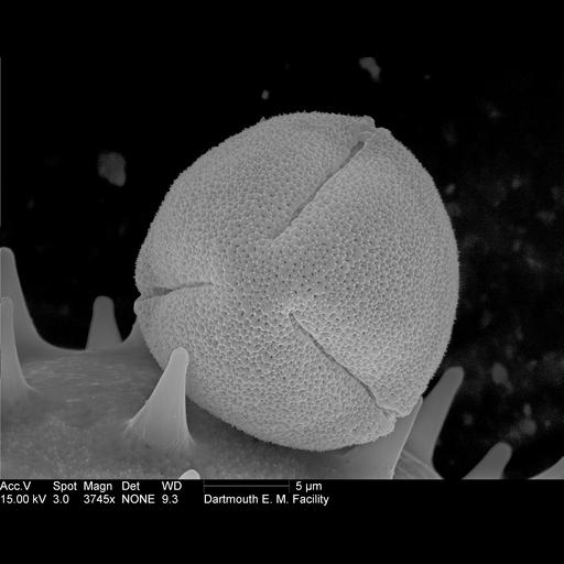
Scanning electron micrograph of Ricinus communis (Castor bean plant) pollen. These specimens have been acetolyzed to remove cytoplasm and pollenkit in order to reveal the intricate wall structure. This is part of a large image collection of scanning electron micrographs of pollen samples. Images of a particular species are grouped together. However, to find images and groups of images from other species, please search for the the name of the contributor, Louisa Howard.

Technical Details

Complete specimen preparation protocol available at http://www.dartmouth.edu/~emlab/manuals/sempreps/pollen.html

Biological Sources
NCBI Organism Classification
Ricinus communis
Cell Type
pollen
Cellular Component
pollen wall
Biological Context
Biological Process
pollen wall assembly
Attribution
Names
Louisa Howard
Link
Dartmouth College EM Facility
Citation
Digital Object Identifier (DOI)
doi:10.7295/W9CIL40284
Archival Resource Key (ARK)
ark:/b7295/w9cil40284
Grouping This image is part of a group.
Imaging
Image Type
recorded image
Image Mode
scanning electron microscopy (SEM)
Parameters Imaged
elastic scattering of electrons
Source of Contrast
differences in deposition of metal shadow
Visualization Methods
gold
Processing History
unprocessed raw data
Sample Preparation
Methods
acetolysis
Relation To Intact Cell
whole mounted tissue
Dimensions
Spatial Axis Image Size Pixel Size
X 3232px ——
Y 2570px ——