Alternate header for print version


Licensing
Public Domain: This image is in the public domain and thus free of any copyright restrictions. However, as is the norm in scientific publishing and as a matter of courtesy, any user should credit the content provider for any public or private use of this image whenever possible. Learn more
tweet  
*CIL – Cell Image Library accession number. Please use this to reference an image.

CIL:40364*  Cite 
Description

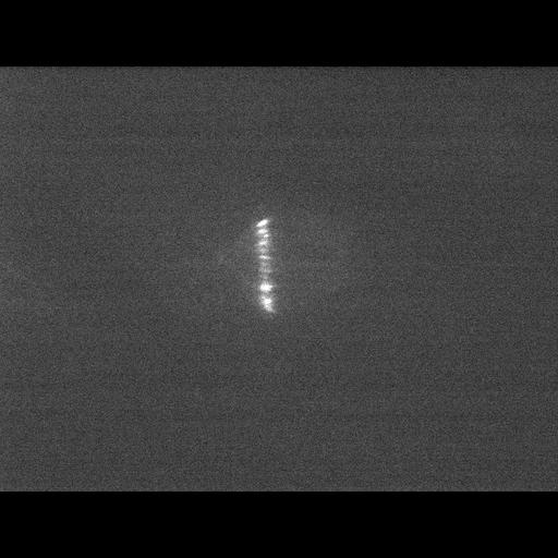
Time series images of a porcine kidney LLCPK1 cell stably expressing photoactivable GFP-tubulin. Fluorescence was activated at metaphase in a vertical strip between the metaphase plate and spindle pole. The activated strip moves poleward, demonstrating a marked microtubule flux during mitotic metaphase. A movie prepared from this time series is included in this group of images.

Technical Details

Cells were imaged using a Nikon Eclipse TE300 microscope with 100x phase NA 1.4 objective lens and spinning disk confocal head (Perkin Elmer) and images captured with a Hamamatsu Orca ER cooled CCD camera every 2 sec. Photoactivation was achieved with a 5-sec exposure to 413-nm light from an X-Cite 120 lamp. See also: NP Ferenz and P Wadsworth 2007. Prophase microtubule arrays undergo flux-like behavior in mammalian cells. Mol Biol Cell 18:3993-4002

Biological Sources
NCBI Organism Classification
Sus scrofa domestica
Cell Type
kidney epithelial cell
Cell Line
LLC-PK1
Cellular Component
microtubule cytoskeleton
Biological Context
Biological Process
mitosis
mitotic metaphase
microtubule cytoskeleton organization
Attribution
Names
Nick Ferenz
Patricia Wadsworth
Citation
Digital Object Identifier (DOI)
doi:10.7295/W9CIL40364
Archival Resource Key (ARK)
ark:/b7295/w9cil40364
Grouping This image is part of a group.
Imaging
Image Type
charge coupled device (CCD)
Image Mode
spinning disk confocal microscopy
Parameters Imaged
fluorescence emission
Source of Contrast
distribution of a specific protein
Visualization Methods
EGFP
Data Qualifiers
processed data
Sample Preparation
Methods
living tissue
Relation To Intact Cell
whole mounted tissue
Dimensions
Spatial Axis Image Size Pixel Size
X 672px ——
Y 512px ——
Time 2 seconds 18