Alternate header for print version


Licensing
Attribution Non-Commercial Share Alike:This image is licensed under a Creative Commons Attribution, Non-Commercial Share Alike License. View License Deed | View Legal Code
tweet  
*CIL – Cell Image Library accession number. Please use this to reference an image.

CIL:40506*  Cite 
Description

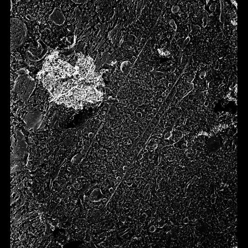
Quick-freeze deep-etch image of rows of carrier vesicles lined up along the cytopharyngeal microtubular ribbons. These vesicles arise from early endosomes as clathrin coated vesicles and are about 100nm in diameter. Vesicles lose their coats before binding with the dynein motors that link them to the microtubules. They tend to lie on the anterior side of the ribbons along with the discoidal vesicles. Carrier vesicles eventually dock on acidosomes and fuse with these. Acidosomes are late endosomes. TEM taken on 5/24/88 by C. Schroeder with Zeiss 10A operating at 80kV. Neg. 12,000X. The raw negative was scanned with an Epson Perfection V750 Pro and this high resolution image is best used for quantitative analysis. Additional information available at (http://www5.pbrc.hawaii.edu/allen/). Small part published in J. Cell Biol. 111:2553-2562, 1990. Adapted with permission.

Biological Sources
NCBI Organism Classification
Paramecium multimicronucleatum
Cell Type
cell by organism
eukaryotic cell
Eukaryotic Protist
Ciliated Protist
Cellular Component
oral apparatus
early endosome membrane
microtubule associated complex
food vacuole
Biological Context
Biological Process
oral apparatus organization
endosome organization
Attribution
Names
Richard Allen (University of Hawaii)
C. Schroeder
Published
J Cell Biol. 1990 Dec;111(6 Pt 1):2553-62.
Pubmed
2148940
Citation
Digital Object Identifier (DOI)
doi:10.7295/W9CIL40506
Archival Resource Key (ARK)
ark:/b7295/w9cil40506
Grouping This image is part of a group.
Imaging
Image Type
transmission electron microscopy (TEM)
illumination by electrons
Image Mode
electron density
Parameters Imaged
elastic scattering of electrons
electron density
elevation
Source of Contrast
differences in deposition of metal shadow
Visualization Methods
transmission electron microscopy (TEM)
Processing History
freeze_fracture/freeze_etch
shadowing and plating
recorded image
film
Data Qualifiers
raw, unprocessed data
suitable for spatial measurements
Dimensions
Spatial Axis Image Size Pixel Size
X 4905px 1.25nm
Y 5350px 1.25nm