Alternate header for print version


Licensing
Attribution Non-Commercial Share Alike:This image is licensed under a Creative Commons Attribution, Non-Commercial Share Alike License. View License Deed | View Legal Code
tweet  
*CIL – Cell Image Library accession number. Please use this to reference an image.

CIL:40554*  Cite 
Description

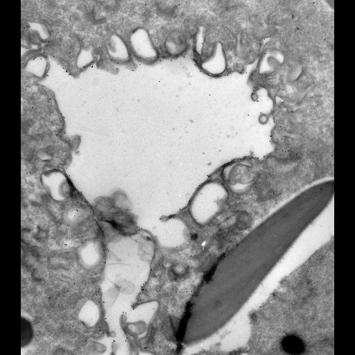
Monoclonal antibody for antigen B2 (called mAb PC-1 in Fok et al., Eur. J. Cell Biol. 40:1-8, 1986) is located on the cytopharynx and along the cytopharyngeal ribbons. It can be seen here on the DV-I membrane that is detached from the cytopharynx. Some acidosomes have already fused with this phagosome. The unfused acidosomes are not labeled with this mAb. TEM taken on 4/3/87 by R. Allen with Zeiss 10A operating at 80kV. Neg. 12,000X. Part published in J. Cell Sci. 108:1263-1274, 1995. Adapted with permission.

Technical Details

To label membranes inside the cell we used very lightly fixed cells (0.25% glutaraldehyde) that were then rapidly frozen in liquid nitrogen and sectioned later at -100oC. These frozen sections were picked up on drops of methylcellulose and transferred to a Formvar-supported grid. The sections were immunogold labeled (15nm gold) to show the location of the specific antigen inside the cell as well as on the cell surface. Microtome sections prepared at approximately 75nm thickness. The raw negative was scanned with an Epson Perfection V750 Pro and this high resolution image is best used for quantitative analysis. Additional information available at (http://www5.pbrc.hawaii.edu/allen/).

Biological Sources
NCBI Organism Classification
Paramecium multimicronucleatum
Cell Type
cell by organism
eukaryotic cell
Eukaryotic Protist
Ciliated Protist
Cellular Component
pre-autophagosomal structure membrane
phagosome
vesicle membrane
Biological Context
Biological Process
digestive system process
Attribution
Names
Richard Allen (University of Hawaii)
Published
J Cell Sci. 1995 Mar;108 ( Pt 3):1263-74.
Pubmed
7622609
Citation
Digital Object Identifier (DOI)
doi:10.7295/W9CIL40554
Archival Resource Key (ARK)
ark:/b7295/w9cil40554
Grouping This image is part of a group.
Imaging
Image Type
transmission electron microscopy (TEM)
illumination by electrons
Image Mode
detection of electrons
film
Parameters Imaged
electron density
intrinsic mass distribution
Source of Contrast
stain with broad specificity
Visualization Methods
stain with broad specificity
uranyl salt
gold
Processing History
recorded image
film
Data Qualifiers
raw, unprocessed data
suitable for spatial measurements
Sample Preparation
Methods
glutaraldehyde fixed tissue
tissue in vitreous ice embedment
microtome-sectioned tissue
monoclonal antibodies
Relation To Intact Cell
microtome-sectioned tissue
Dimensions
Spatial Axis Image Size Pixel Size
X 4999px 1.25nm
Y 5660px 1.25nm