Alternate header for print version


Licensing
This image is copyright protected. Any public or private use of this image is subject to prevailing copyright laws. Please contact the content provider of this image for permission requests.
tweet  
*CIL – Cell Image Library accession number. Please use this to reference an image.

CIL:40625*  Cite 
Description

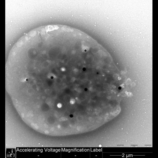
Transmission electron micrograph of unfixed whole mounted platelet upon an EM support film from platelet rich plasma. The dense granules contain calcium rendering them electron opaque. The “dark” granules are delta granules within the platelet cytoplasm (contain calcium, adenine nucleotides, serotonin and pryrophosphates), and the bright white circles likely represent a dilated open canalicular system.

Technical Details

Image collected on an FEI Tecnai G2 Spirit transmission electron microscope.

Biological Sources
NCBI Organism Classification
Homo sapiens
Cell Type
platelet
Cellular Component
cell surface
platelet dense granule
platelet delta granules
Attribution
Names
William T. Gunning III
OTHER
Pathology Department, University of Toledo
Citation
Digital Object Identifier (DOI)
doi:10.7295/W9CIL40625
Archival Resource Key (ARK)
ark:/b7295/w9cil40625
Imaging
Image Type
recorded image
Image Mode
transmission electron microscopy (TEM)
Parameters Imaged
electron density
Visualization Methods
visualization of contiguous regions
Processing History
unprocessed raw data
Sample Preparation
Methods
unfixed tissue
Relation To Intact Cell
whole mounted tissue
Dimensions
Spatial Axis Image Size Pixel Size
X 2048px ——
Y 2198px ——