Alternate header for print version


Licensing
Attribution Non-Commercial; No Derivatives:This image is licensed under a Creative Commons Attribution, Non-Commercial, No Derivatives License. View License Deed | View Legal Code
tweet  
*CIL – Cell Image Library accession number. Please use this to reference an image.

CIL:40965*  Cite 
Description

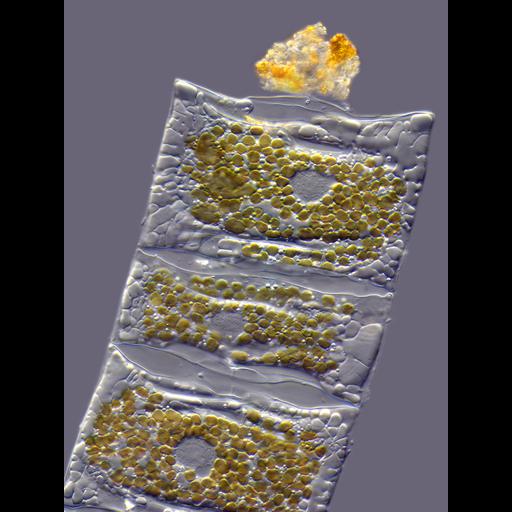
Brightfield micrograph of a living diatom (Mediopyxis helysia) showing the cell nuclei and golden chloroplasts. There is a bacteria colony in mucilage located on the top of the diatom. The image won the Ninth Prize, 2011 Olympus BioScapes Digital Imaging Competition®.

Technical Details

Biological Sources
NCBI Organism Classification
Mediopyxis helysia
Cellular Component
nucleus
chloroplast
Biological Context
Biological Process
organelle organization
Attribution
Names
Wolfgang Bettighofer
2011 Olympus BioScapes Digital Imaging Competition®
Link
2011 Olympus BioScapes Digital Imaging Compe...
OTHER
Kiel, Germany
Citation
Digital Object Identifier (DOI)
doi:10.7295/W9CIL40965
Archival Resource Key (ARK)
ark:/b7295/w9cil40965
Imaging
Image Type
recorded image
Image Mode
bright-field microscopy
Parameters Imaged
absorption of illumination
Source of Contrast
differences in intrinsic optical density
Visualization Methods
visualization of contiguous regions
Sample Preparation
Methods
living tissue
Relation To Intact Cell
whole mounted tissue
Dimensions
Spatial Axis Image Size Pixel Size
X 2304px ——
Y 3072px ——