Alternate header for print version


Licensing
Attribution Non-Commercial Share Alike:This image is licensed under a Creative Commons Attribution, Non-Commercial Share Alike License. View License Deed | View Legal Code
tweet  
*CIL – Cell Image Library accession number. Please use this to reference an image.

CIL:40994*  Cite 
Description

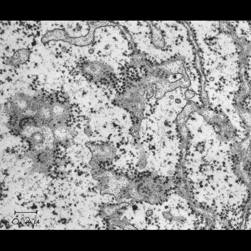
Transmission electron micrograph of a thin section of the liver of a 1-day old rat showing a grazing view of rough endoplasmic reticulum (RER, sometimes termed granular endoplasmic reticulum. The 'rough' or 'granular' term refers to the membrane-attached polyribosomes (also known as polysomes) which often occur in a spiral or rosettal conformation. RER-associated polyribosomes are the sites of synthesis of proteins secreted into the RER cisternae and destined for export from the cell.

Technical Details

Original 3.25 in. x 4 in. lantern slides were scanned at 600dpi.

Biological Sources
NCBI Organism Classification
Rattus rattus
Cell Type
hepatocyte
Cellular Component
rough endoplasmic reticulum
polysome
Biological Context
Biological Process
protein synthesis
cytoplasm organization
endoplasmic reticulum organization
Attribution
Names
George E. Palade
Link
George E. Palade EM Slide Collection
Citation
Digital Object Identifier (DOI)
doi:10.7295/W9CIL40994
Archival Resource Key (ARK)
ark:/b7295/w9cil40994
Grouping This image is part of a group.
Imaging
Image Type
film
Image Mode
transmission electron microscopy (TEM)
Parameters Imaged
electron density
Source of Contrast
differences in adsorption or binding of stain
Visualization Methods
uranyl salt
osmium tetroxide
lead salt
Data Qualifiers
processed data
Sample Preparation
Methods
chemically fixed tissue
tissue in plastic embedment
Relation To Intact Cell
microtome-sectioned tissue
Dimensions
Spatial Axis Image Size Pixel Size
X 4046px ——
Y 3382px ——