Alternate header for print version


Licensing
Attribution Non-Commercial Share Alike:This image is licensed under a Creative Commons Attribution, Non-Commercial Share Alike License. View License Deed | View Legal Code
tweet  
*CIL – Cell Image Library accession number. Please use this to reference an image.

CIL:40997*  Cite 
Description

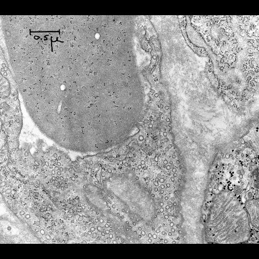
Transmission electron micrograph of a thin section of a capillary from the ventricular myocardium of the rat showing part of a reticulocyte (immature erythrocyte) in the lumen of the capillary. The reticulocyte cytoplasm is devoid of membranous organelles but contains free polyribosomes (also known as polysomes) which primarily synthesize globin during the late stages of erythrocyte maturation.

Technical Details

Original 3.25 in. x 4 in. lantern slides were scanned at 600dpi.

Biological Sources
NCBI Organism Classification
Rattus rattus
Cell Type
reticulocyte
Cell Line
Endothelium of capillary
Cellular Component
polysome
cytoplasm
Biological Context
Biological Process
protein synthesis
cytoplasm organization
Attribution
Names
George E. Palade
Link
George E. Palade EM Slide Collection
Citation
Digital Object Identifier (DOI)
doi:10.7295/W9CIL40997
Archival Resource Key (ARK)
ark:/b7295/w9cil40997
Grouping This image is part of a group.
Imaging
Image Type
film
Image Mode
transmission electron microscopy (TEM)
Parameters Imaged
electron density
Source of Contrast
differences in adsorption or binding of stain
Visualization Methods
uranyl salt
osmium tetroxide
lead salt
Data Qualifiers
processed data
Sample Preparation
Methods
chemically fixed tissue
tissue in plastic embedment
Relation To Intact Cell
microtome-sectioned tissue
Dimensions
Spatial Axis Image Size Pixel Size
X 3805px ——
Y 3364px ——