Alternate header for print version


Licensing
Attribution Non-Commercial Share Alike:This image is licensed under a Creative Commons Attribution, Non-Commercial Share Alike License. View License Deed | View Legal Code
tweet  
*CIL – Cell Image Library accession number. Please use this to reference an image.

CIL:41043*  Cite 
Description

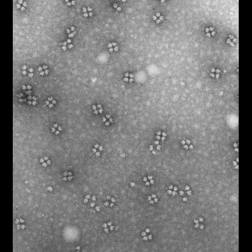
Transmission electron micrograph of a negatively stained preparation of ribosomes isolated from a 3-day old chick embryo that was maintained in a cold room for 24 hrs. The ribosome crystals seen in thin sections of these cells (see CIL:41041)are seen to be primarily tetramers.

Technical Details

See: T. Morimoto et al. 1972. Ribosome crystallization in chicken embryos. I. Isolation, characterization, and in vitro activity of ribosome tetramers. J Cell Biol 52:355-366. B. Byers 1966. Ribosome crystallization induced in chick embryo tissues by hypothermia. J Cell Biol 30:C1-6. Original 3.25 in. x 4 in. lantern slides were scanned at 600dpi

Biological Sources
NCBI Organism Classification
Mus musculus
Cell Type
early embryonic cell
Cellular Component
ribosome
Biological Context
Biological Process
protein synthesis
cytoplasm organization
ribosome biogenesis
Attribution
Names
George E. Palade
Link
George E. Palade EM Slide Collection
Citation
Digital Object Identifier (DOI)
doi:10.7295/W9CIL41043
Archival Resource Key (ARK)
ark:/b7295/w9cil41043
Grouping This image is part of a group.
Imaging
Image Type
film
Image Mode
transmission electron microscopy (TEM)
Parameters Imaged
electron density
Source of Contrast
differences in adsorption or binding of stain
Visualization Methods
negative staining
Data Qualifiers
processed data
Sample Preparation
Relation To Intact Cell
isolated subcellular component
Dimensions
Spatial Axis Image Size Pixel Size
X 3381px ——
Y 3770px ——