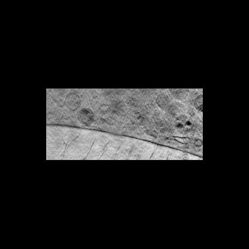
The image shows a portion of a slice through a 3D tomographic reconstruction of a mouse adenocarcinoma cell based on x-ray microscopy. Live cells were cryofixed by plunge freezing in liquid ethane and the vitrified material observed unstained using x-ray microscopy at a wavelength that yields high contrast between water and organic material. Images were taken over a range of specimen tilts from -60 to +60 degrees at 1 degree intervals. The image represents a 9.8 nm thick slice of the computed tomogram in the x-y plane, and shows the plasma membrane with cytoplasm above and medium below. Numerous vesicles are present in the cytoplasm. The image has a resolution of ~36 nm (Rayleigh Criterion) and ~70 nm (Fourier Ring Correlation), the latter corresponding to ~35 nm half-pitch resolution. Additional X-ray images from the study are included in this CIL Group. See also: W G Muller et al. 2012 Towards an atlas of mammalian cell ultrastructure by soft X-ray tomography. J Struct Biol 177:179-192.
Mouse adenocarcinoma cells were grown on gold grids coated with cellulose nitrate, incubated with 270 nm diameter gold-coated silica beads, plunge frozen in liquid ethane, transferred to a Gatan 630 cryoholder, and imaged at -170C in the frozen hydrated state using the X-ray microscope at the Helmholz Zentrum, Berlin, Germany. The microscope uses partially coherent illumination and a zone plate objective, and allows the specimen to be observed over a range of tilts. The images were aligned using the gold beads as fiduciary markers, and reconstructed using a reciprocal space algorithm. See: G Schneider et al. 2010 Three-dimensional cellular ultrastructure resolved by X-ray microscopy. Nat Meth 7:985-987.
| Spatial Axis | Image Size | Pixel Size |
|---|---|---|
| X | 322px | 19.6nm |
| Y | 147px | 19.6nm |