Alternate header for print version


Licensing
Attribution Non-Commercial; No Derivatives:This image is licensed under a Creative Commons Attribution, Non-Commercial, No Derivatives License. View License Deed | View Legal Code
tweet  
*CIL – Cell Image Library accession number. Please use this to reference an image.

CIL:41558*  Cite 
Description

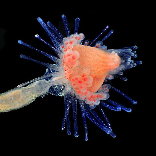
Hydroid collected from kelp sample captured using epi-illumination and an image stack. Honorable Mention, 2011 Olympus BioScapes Digital Imaging Competition®.

Technical Details

Biological Sources
NCBI Organism Classification
hydroid
Cellular Component
cell surface
Biological Context
Biological Process
polyp stage of life-cycle
Attribution
Names
Mike Crutchley
2011 Olympus BioScapes Digital Imaging Competition®
Link
2011 Olympus BioScapes Digital Imaging Compet...
OTHER
Pembrokeshire, Wales, United Kingdom
Citation
Digital Object Identifier (DOI)
doi:10.7295/W9CIL41558
Archival Resource Key (ARK)
ark:/b7295/w9cil41558
Imaging
Image Type
recorded image
Image Mode
bright-field microscopy
epi-illumination
Parameters Imaged
absorption of illumination
Source of Contrast
differences in intrinsic optical density
Processing History
assembly of image stack
Sample Preparation
Relation To Intact Cell
whole mounted tissue
Dimensions
Spatial Axis Image Size Pixel Size
X 3384px ——
Y 2848px ——