Alternate header for print version


Licensing
Attribution Non-Commercial; No Derivatives:This image is licensed under a Creative Commons Attribution, Non-Commercial, No Derivatives License. View License Deed | View Legal Code
tweet  
*CIL – Cell Image Library accession number. Please use this to reference an image.

CIL:41658*  Cite 
Description

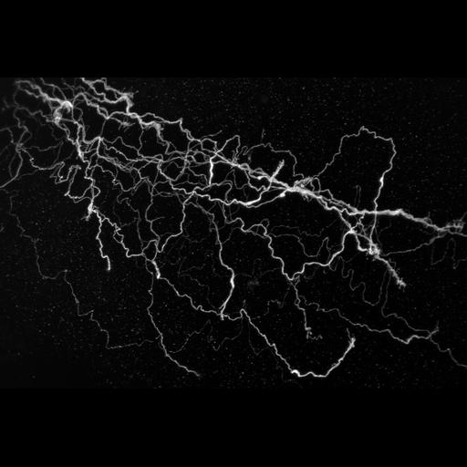
Confocal micrograph of “lost” axons - chick interneurons labeled with green fluorescent protein that were transplanted from older into younger embryos. When they encounter unfamiliar territory, the axons create these trails as they try to recognize their pre-programmed trajectory. This image was first published in Developmental Biology 297, 508-21. Honorable Mention, 2011 Olympus BioScapes Digital Imaging Competition®.

Technical Details

Biological Sources
NCBI Organism Classification
Gallus gallus
Cell Type
interneuron
Cellular Component
neuron projection
Biological Context
Biological Process
axon guidance
Attribution
Names
Richard Wingate
2011 Olympus BioScapes Digital Imaging Competition®
Link
2011 Olympus BioScapes Digital Imaging Compe...
OTHER
MRC Center for Developmental Neurobiology, King’s College London, England, UK
Citation
Digital Object Identifier (DOI)
doi:10.7295/W9CIL41658
Archival Resource Key (ARK)
ark:/b7295/w9cil41658
Imaging
Image Type
recorded image
Image Mode
confocal microscopy
Parameters Imaged
fluorescence emission
Source of Contrast
distribution of a specific protein
Visualization Methods
EGFP
Sample Preparation
Relation To Intact Cell
whole mounted tissue
Dimensions
Spatial Axis Image Size Pixel Size
X 768px ——
Y 512px ——