Alternate header for print version


Licensing
Attribution Non-Commercial; No Derivatives:This image is licensed under a Creative Commons Attribution, Non-Commercial, No Derivatives License. View License Deed | View Legal Code
tweet  
*CIL – Cell Image Library accession number. Please use this to reference an image.

CIL:41734*  Cite 
Description

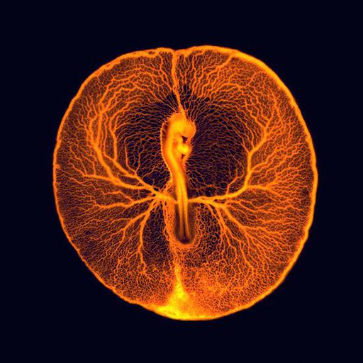
This image of the vascular system of a chicken embryo taken two days after fertilization is a composite of two different images taken with an upright fluorescent dissecting scope. The egg's shell was opened and fluorescent dextran was injected into the lower part of the vascular system. The heart pumped the dye and revealed the entire vasculature used by the embryo to feed itself from the rich underlying yolk inside the egg. Wellcome Image Award 2012.

Technical Details

B0008295. 2011 Collection: Wellcome Images Copyrighted work available under Creative Commons by-nc-nd 2.0 UK: England & Wales, see http://images.wellcome.ac.uk/indexplus/page/Prices.html

Biological Sources
NCBI Organism Classification
Gallus gallus
Biological Context
Biological Process
vasculogenesis
embryo development
Attribution
Names
Vincent Pasque
Link
Wellcome Images
OTHER
University of Cambridge
Citation
Digital Object Identifier (DOI)
doi:10.7295/W9CIL41734
Archival Resource Key (ARK)
ark:/b7295/w9cil41734
Imaging
Image Type
recorded image
Image Mode
fluorescence microscopy
Parameters Imaged
fluorescence emission
Source of Contrast
compartmentalization of stain or label
Processing History
composite of two images
Sample Preparation
Relation To Intact Cell
whole mounted tissue
Dimensions
Spatial Axis Image Size Pixel Size
X 550px ——
Y 550px ——