Alternate header for print version


Licensing
Attribution Non-Commercial; No Derivatives:This image is licensed under a Creative Commons Attribution, Non-Commercial, No Derivatives License. View License Deed | View Legal Code
tweet  
*CIL – Cell Image Library accession number. Please use this to reference an image.

CIL:41736*  Cite 
Description

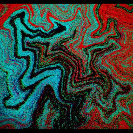
A confocal micrograph showing Bacillus subtilis, a Gram-positive, rod-shaped bacterium that is commonly found in soil. Distinct lineages of bacteria expressing different fluorescent proteins were initially mixed randomly on a petri dish. As the bacteria grow, they organize themselves into reproducible patterns and shapes that can be predicted with mathematical models. This image is part of a synthetic biology project involved in designing artificial genetic circuits for pattern formation in bacterial colonies and plant tissues. The horizontal field width of this image is 500 microns. Wellcome Image Award 2012.

Technical Details

B0008257. 2011 Collection: Wellcome Images Copyrighted work available under Creative Commons by-nc-nd 2.0 UK: England & Wales, see http://images.wellcome.ac.uk/indexplus/page/Prices.html

Biological Sources
NCBI Organism Classification
Bacillus subtilis
Cellular Component
cell surface
Biological Context
Biological Process
biofilm organization
Attribution
Names
Fernan Federici
Jim Haseloff
Citation
Digital Object Identifier (DOI)
doi:10.7295/W9CIL41736
Archival Resource Key (ARK)
ark:/b7295/w9cil41736
Imaging
Image Type
recorded image
Image Mode
confocal microscopy
Parameters Imaged
fluorescence emission
Source of Contrast
distribution of epitope
Visualization Methods
TagRFP-T
sfGFP
mTagBFP
mKate2FP
mOrange2FP
Sample Preparation
Relation To Intact Cell
dispersed cells in vitro
Dimensions
Spatial Axis Image Size Pixel Size
X 583px ——
Y 550px ——