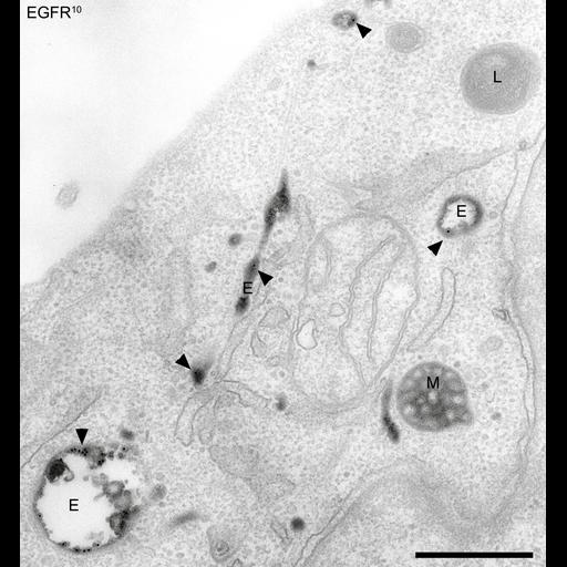
Transmission electron micrograph of a thin section of a HeLa cell treated to reveal components of the endocytic pathway. Epidermal growth factor (EGFR) is labeled with 10 nm immuno-gold particles and the localization of transferrin (Tf) is seen as an electron dense product as a result of treatment with Tf conjugated to horse radish peroxidase followed by diamino benzidine. E, early endosome M, late endosome (multivesicular body) L, lysozome Arrowheads denote gold particles Scale bar 500 nm
Epidermal growth factor receptors were labelled with an antibody conjugated to 10 nm gold, applied to the cell surface of HeLa cells, and stimulated with EGF, which causes internalization of the receptors and attached gold. The image was taken 5 minutes after internalization, at which point most of the EGFR-gold has reached early endosomes (black dots, marked by arrowheads), but has not yet entered late endosomes or lysosomes. The cells were also loaded with transferrin conjugated to horseradish peroxidase (TfHRP). The HRP catalyses a reaction in the presence of DAB that produces an electron dense product in the transferrin-containing compartments in the image. After a 1h preincubation in serum-free medium, cells were exposed to TfHRP and then incubated with EGF and anti-EGFR 10 nm gold-conjugated antibody for 30 minutes at 4C, washed, and incubated at 37C for a further 5 minutes, all in the presence of TfHRP. Cells were then room temperature fixed in 2% PFA/1.5% glutaraldehyde, a DAB reaction was carried out, and the cells were osmium stained, tannic acid treated, dehydrated, embedded en face in epon, and ~70 nm sections were cut and observed in a Philips 400 TEM. Images were recorded on film and subsequently digitized with a Flextight Precision II scanner (Imacon). Images were processed for brightness/contrast and annotations in Adobe Photoshop 5.0 See also: Doyotte, A., Russell, M.R.G., Hopkins, C.R., Woodman, P.G. (2005) Depletion of TSG101 forms a mammalian "Class E" compartment: a multicisternal early endosome with multiple sorting defects. J. Cell Sci, 118:3003-3017
| Spatial Axis | Image Size | Pixel Size |
|---|---|---|
| X | 3259px | —— |
| Y | 3510px | —— |