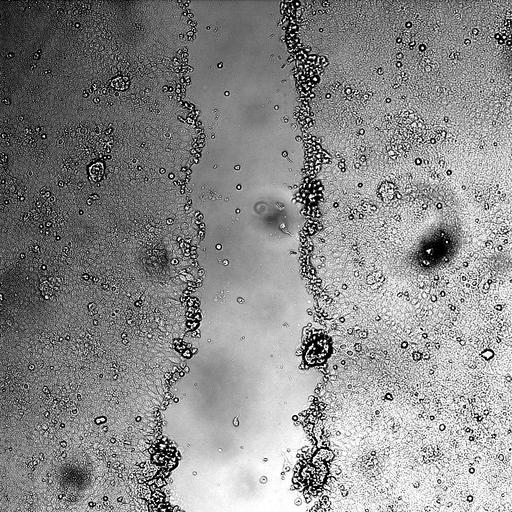
Time series light microscopy images illustrating a wound healing assay. A monolayer of MDCK (Madin-Darby Canine Kidney) epithelial cells is scratched to create a 'wound' about 300 micrometers in width, after which the growth of the cells to fill the wound is monitored by recording images over the course of 15h. CIL:44511 contains a movie of the process. This image is part of a group of 12 that includes control cells (CIL:44501, 502, 506, 507, 508) and after addition of Hepatocyte Growth Factor /Scatter Factor (HGF/SF) to activate HGF/SF-Met signaling (CIL:44503, 504, 505, 509, 510). Also in the group are two movies, one control (CIL:44511), and one HGF/SF treated (CIL:44512). For additional wound healing assays see CIL:43401 and images grouped with it.
Madin-Darby Canine Kidney (MDCK) epithelial cells expressing YFP-membrane were maintained in DMEM supplemented with 5% fetal FCS in a 37°C, 5% CO2 incubator. Wound healing assay: Cells were grown to 90% confluence in 24-well plates. Prior to scratching, the cells were starved by changing the medium to DMEM plus 0.1% FCS (starvation medium) for 24 hours. The medium was then changed to either fresh starvation medium (control), or starvation medium with 80 ng ml-1 HGF/SF for an additional 2 hours. A scratch of approximately 300 μm in width was generated using a 200 μl tip. The plate was subjected to time lapse microscopy in a stage incubator (OKOLAB, Italy) on a computer-controlled motorized stage of a confocal microscope (CLSM-510, Carl Zeiss, Germany), used in non-confocal DIC mode, with a 10x (NA 0.30) objective. Image acquisition was initiated 2 hours post scratching. Images were acquired every 15.7 minutes for 15 hours. The coordinates of each scratch were predefined, and a macro that repetitively positions the field of view at each point was executed. The acquired differential interference contrast (DIC) channel of the time-lapse sequence was used for the analysis.
| Spatial Axis | Image Size | Pixel Size |
|---|---|---|
| X | 1024px | 0.879µm |
| Y | 1024px | 0.879µm |
| Time | 942 seconds | 60 |
|---|